

Microscope: Complete Master Guide
History · Types (16+ explained) · Parts & Functions · Optical Principles · Step-by-Step Procedures · Staining · Maintenance · Advanced Microscopy & Applications

📌 What is a Microscope?
Derived from Greek "mikros" (small) and "skopein" (to look). It is a precision instrument designed to magnify small objects that cannot be seen by the naked eye.
Microscopy is the technical field of using microscopes to view samples and objects that cannot be seen with the unaided eye (objects that are not within the resolution range of the normal eye).
💡 The Resolution Threshold
The human eye can resolve objects down to 0.1mm (100 micrometers). Anything smaller, such as cells (1-100 μm) or viruses (20-300 nm), requires mechanical magnification.
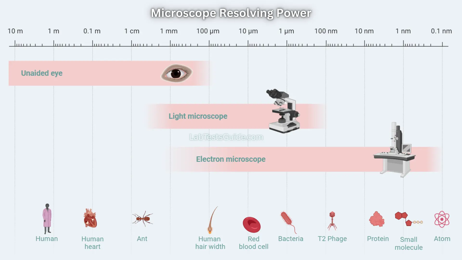
History & Evolution of Microscopy
🔎 Ancient & Early Beginnings
Assyrians (700 BC) used lenses. Romans employed glass spheres. In 1590, Dutch spectacle makers Zacharias & Hans Janssen created first compound microscope — a tube with lenses. Marked the dawn of microbiology.
🔬 Golden Age: 17th Century
Antonie van Leeuwenhoek (1632-1723) perfected single-lens microscopes (275x), discovered bacteria, sperm cells, blood cells. Robert Hooke published Micrographia (1665) — coined the term "cell".
16 Types of Microscopes – Complete Classification
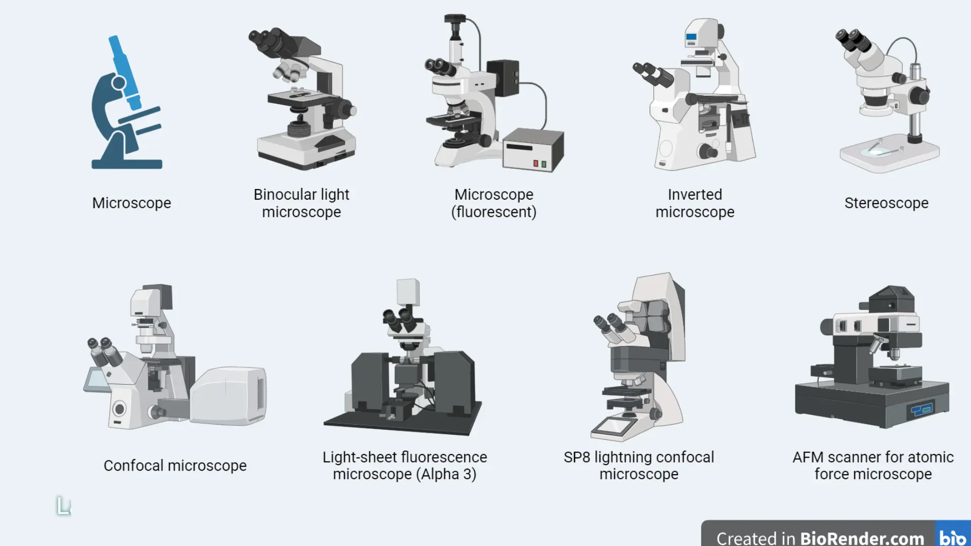
| Type | Principle / Key Feature | Magnification | Primary Applications |
|---|---|---|---|
| Simple Microscope | Single convex lens (magnifying glass) | 5x – 50x | Jewelry inspection, basic education, watchmaking |
| Compound Microscope | Two lens systems (objective + eyepiece), brightfield | 40x – 1000x (2000x oil) | Cell biology, pathology, microbiology, routine lab |
| Stereo (Dissecting) | Two optical paths, 3D image, low magnification | 10x – 80x | Dissection, entomology, circuit board inspection |
| Phase-Contrast | Converts phase shifts to contrast (live cells) | 100x – 1000x | Observing unstained living cells, sperm motility |
| Fluorescence | UV/blue light excites fluorophores | 100x – 1000x | Protein localization, immunofluorescence, FISH |
| Confocal (Laser Scanning) | Pinhole eliminates out-of-focus light, optical sectioning | 100x – 1000x+ | 3D reconstruction of thick specimens, live cell imaging |
| Polarizing | Polarized light with analyzer; detects birefringence | 40x – 1000x | Geology (minerals), polymer stress analysis |
| Dark Field | Direct light blocked; only scattered light from specimen | 40x – 1000x | Viewing spirochetes, unstained flagella, live bacteria |
| Inverted Microscope | Objectives below stage, light from above | 100x – 400x | Live cell culture (Petri dishes, flasks), IVF |
| Digital Microscope | Camera sensor replaces eyepiece; image on screen | 20x – 1000x | Teaching, documentation, quality control |
| USB Microscope | Portable, connects to computer | 20x – 250x | Field work, hobbyists, quick inspection |
| Transmission EM (TEM) | Electrons transmitted through ultra-thin sample | Up to 10,000,000x | Internal ultrastructure (viruses, organelles, proteins) |
| Scanning EM (SEM) | Electron beam scans surface; 3D topography | 10x – 500,000x | Surface morphology, insects, fractures, nanomaterials |
| Atomic Force Microscope (AFM) | Cantilever tip scans surface (intermolecular forces) | Atomic resolution | Nanotechnology, molecular biophysics, polymers |
| Scanning Tunneling (STM) | Tunneling current between tip and conductive surface | Atomic | Surface atoms of conductive materials |
| Acoustic / X-Ray Microscope | Sound waves or X-rays for imaging | Variable | Non-destructive testing, thick biological specimens |
📌 Emerging Trends 2026
AI-powered automated microscopy, deep-learning denoising, expansion microscopy (physical specimen expansion), and MINFLUX nanoscopy achieving 1 nm resolution in living cells.
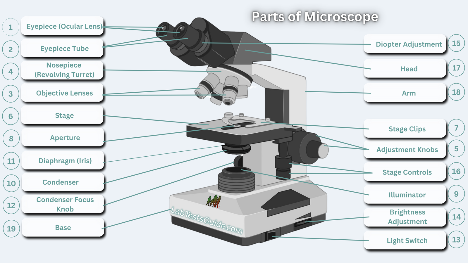
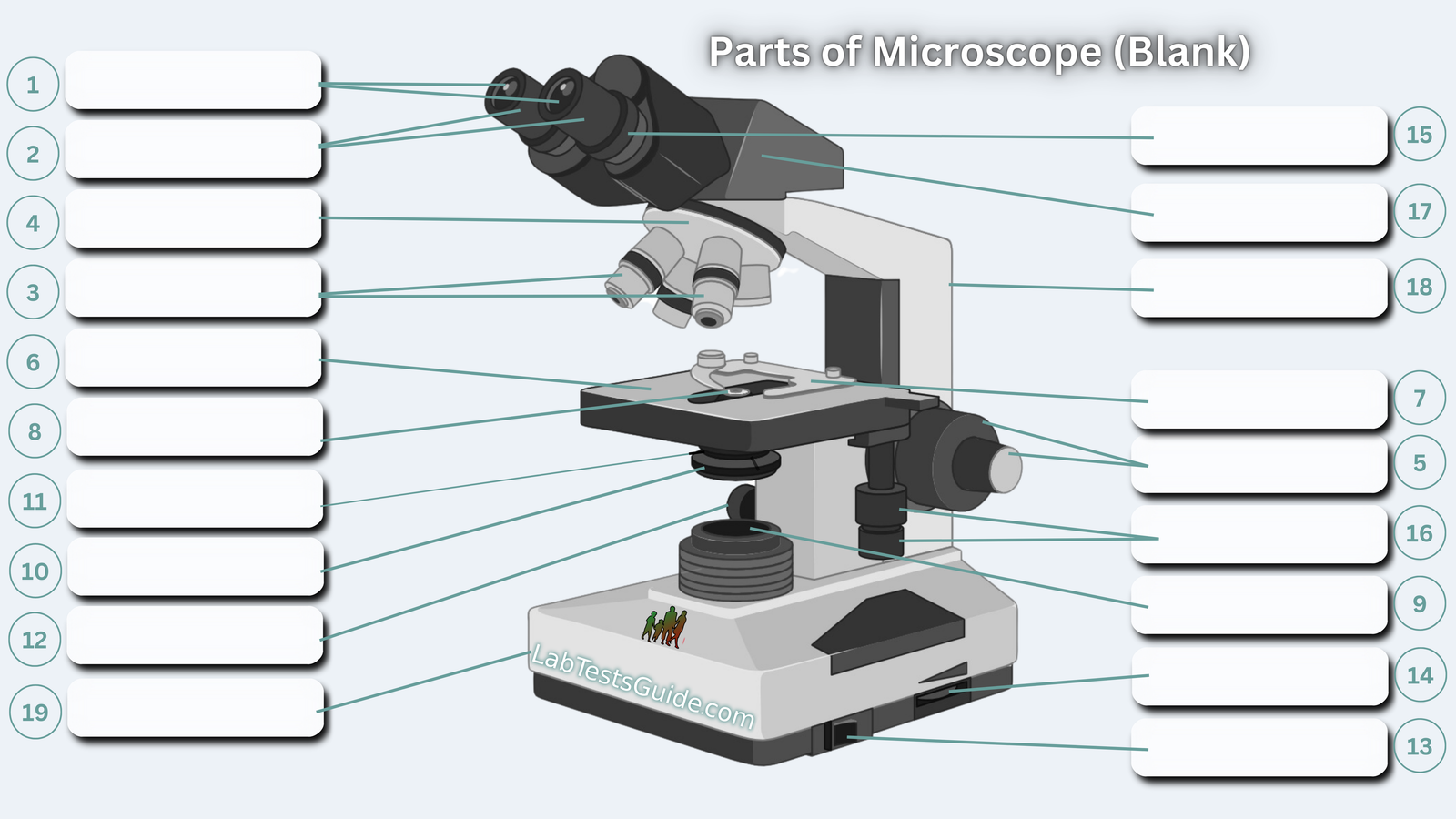
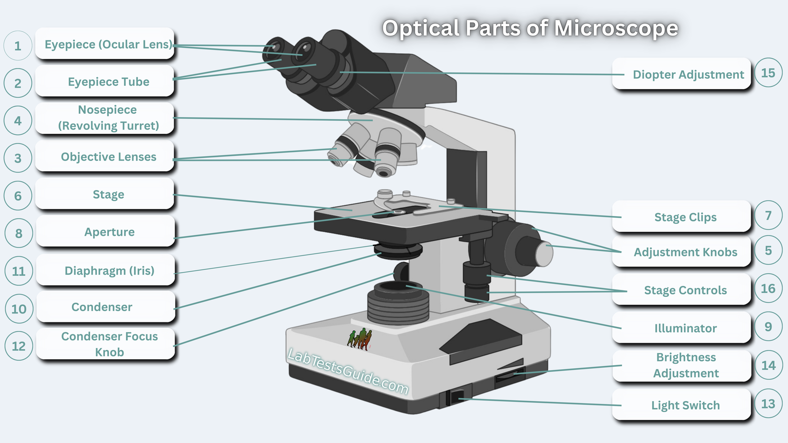
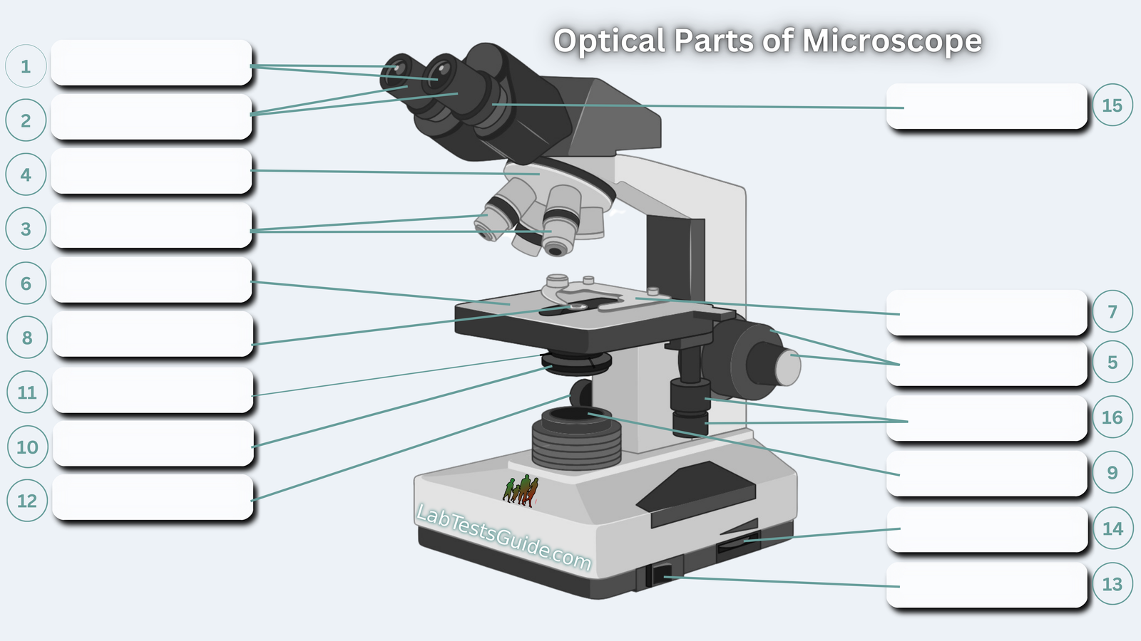
Anatomy of a Microscope – 19 Essential Parts & Functions
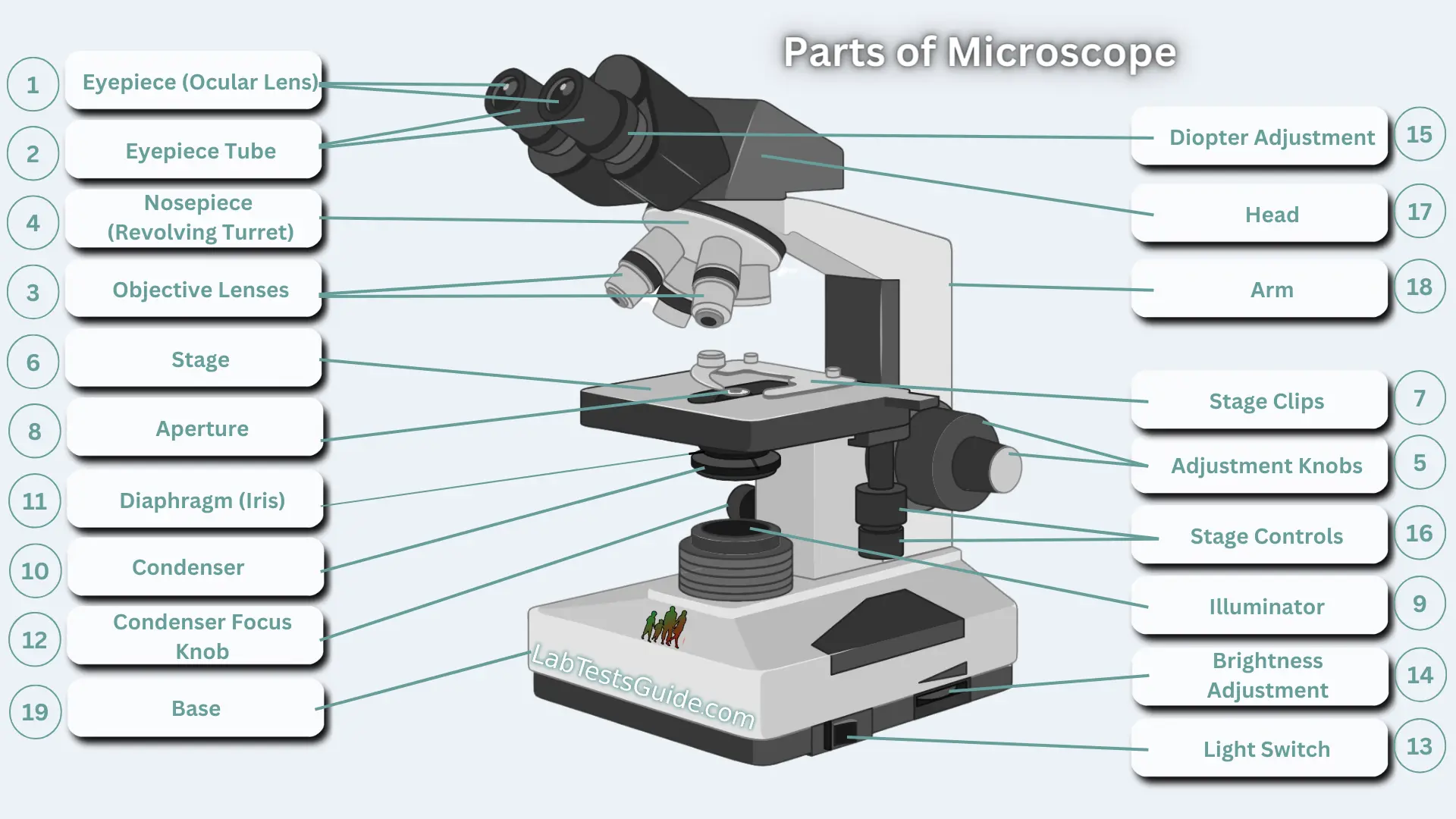
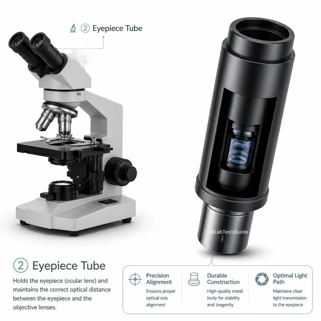
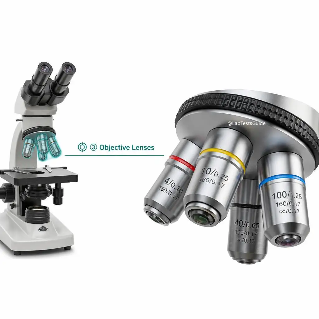
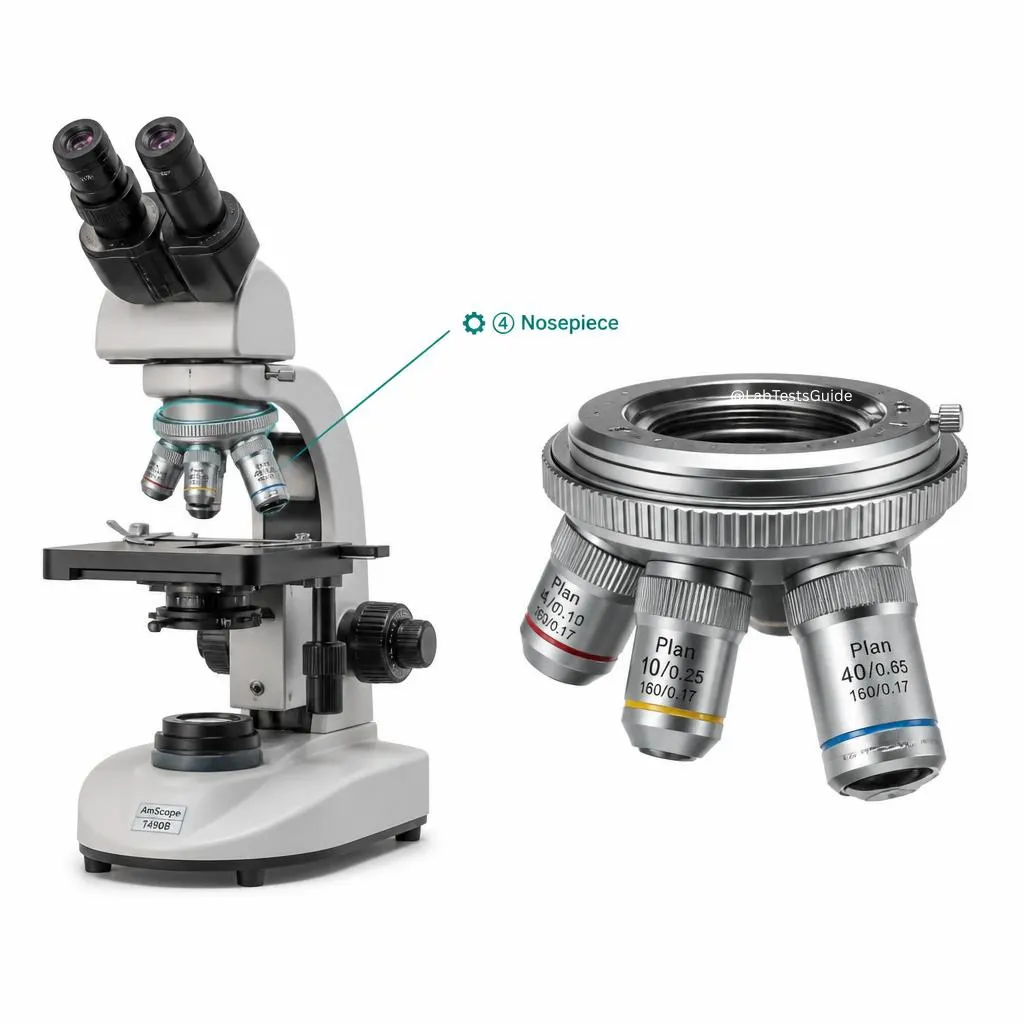
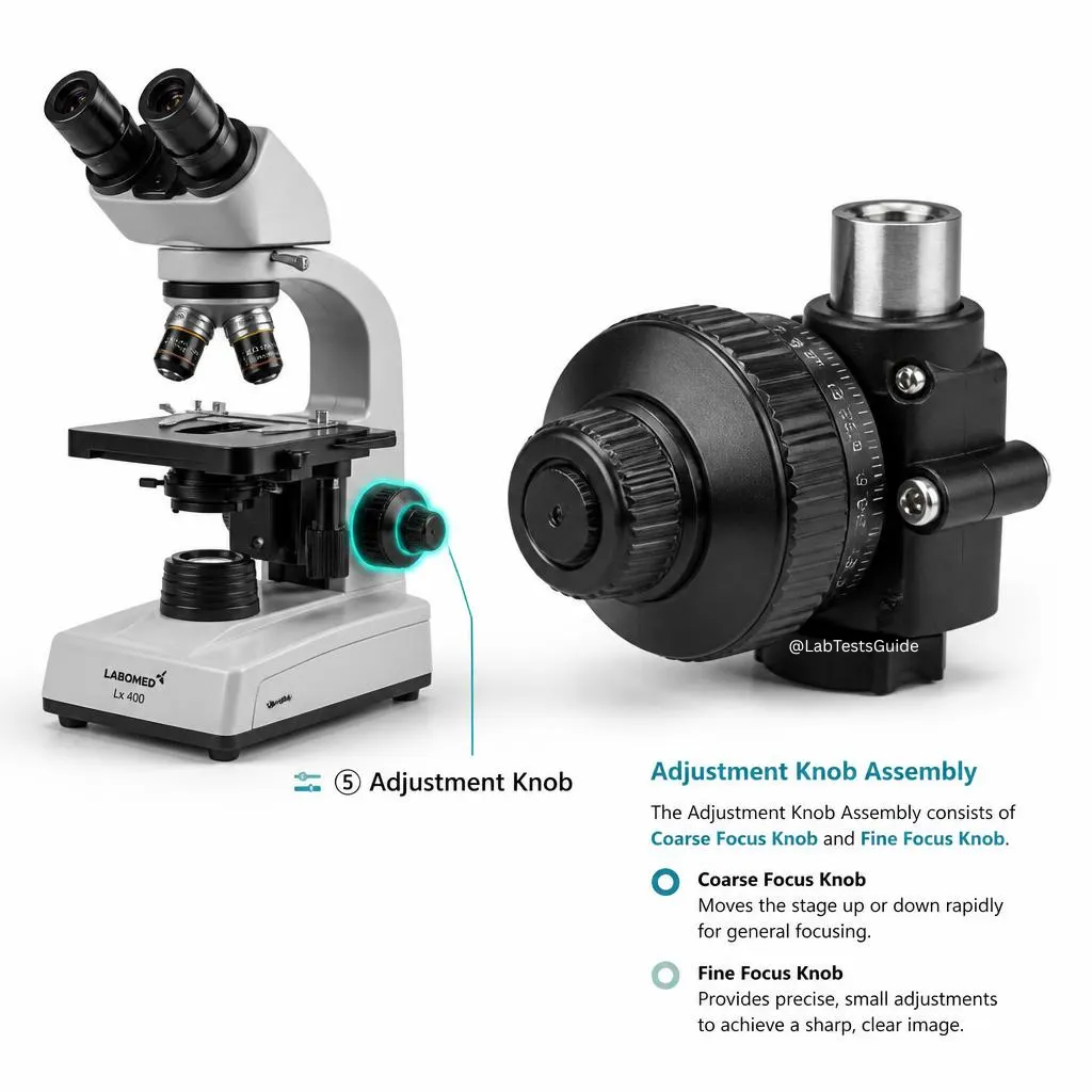
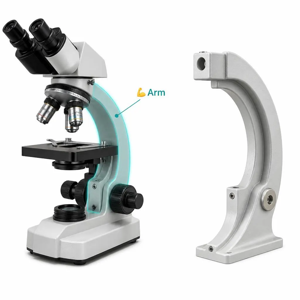
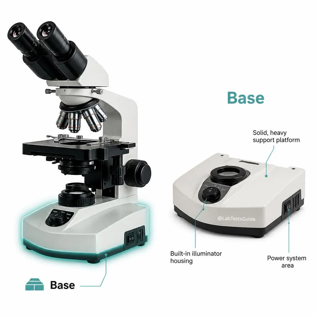
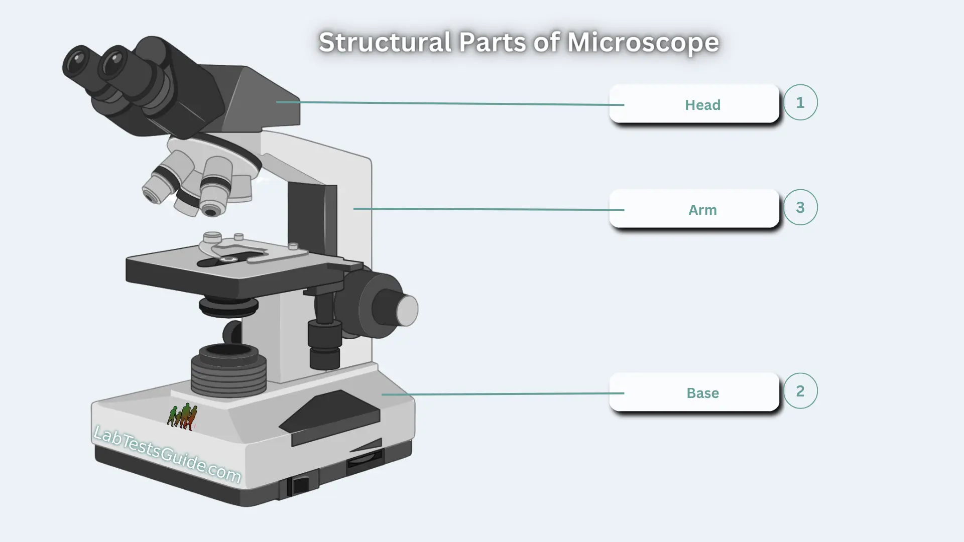
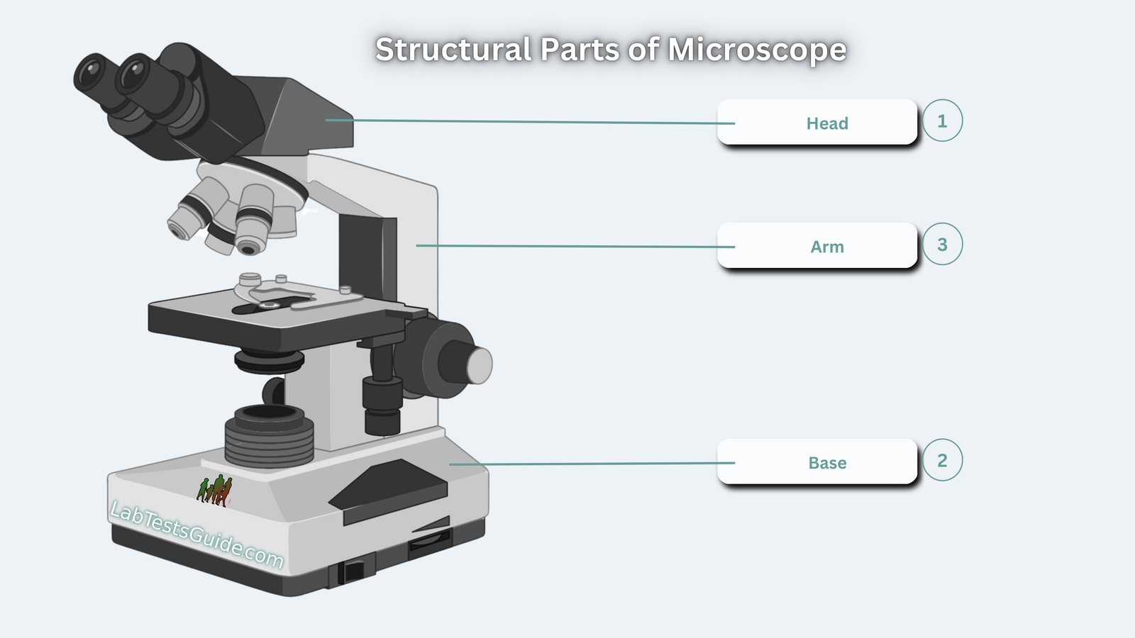
Structural parts
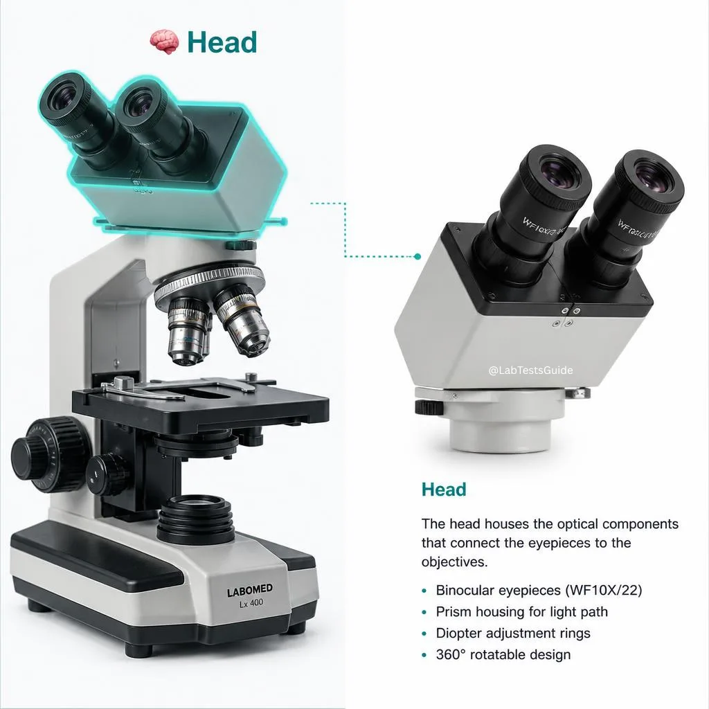
- Head (Body tube): holds eyepiece and objectives
- Arm: curved support for carrying
- Base: stable foundation, houses illuminator
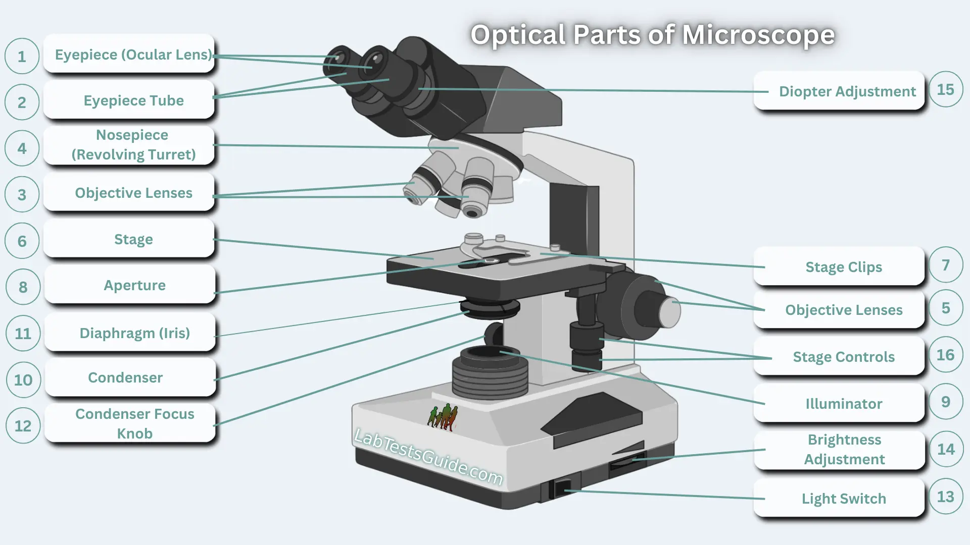
Optical & adjustment parts
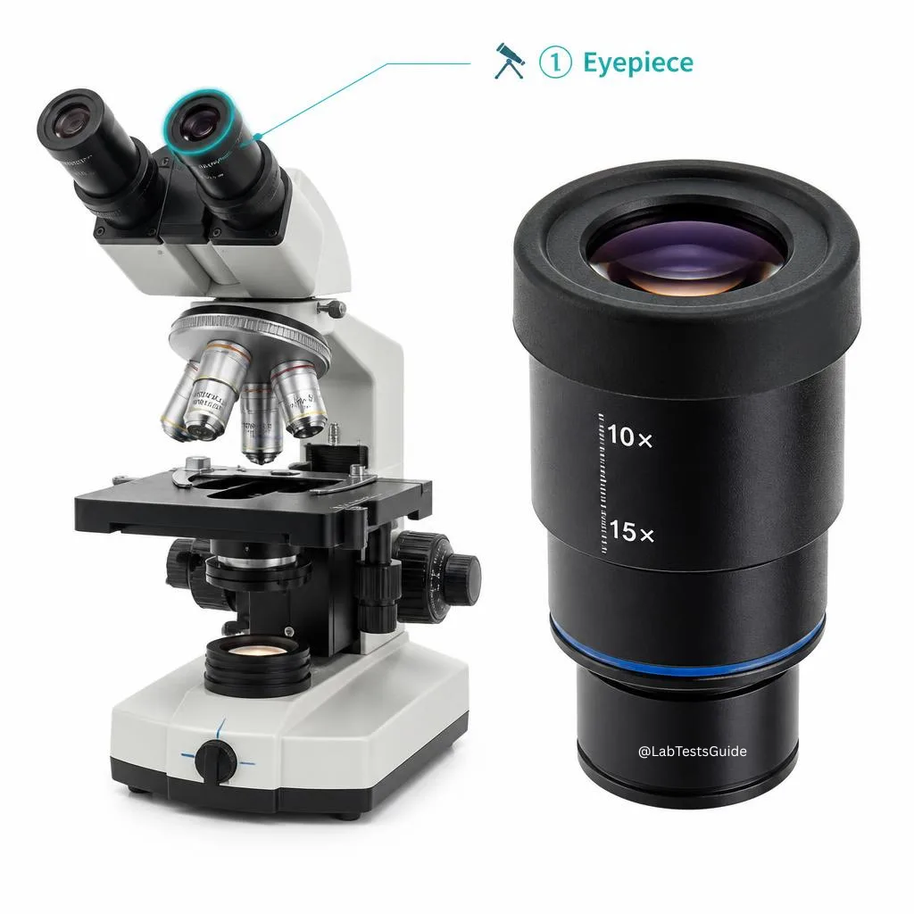
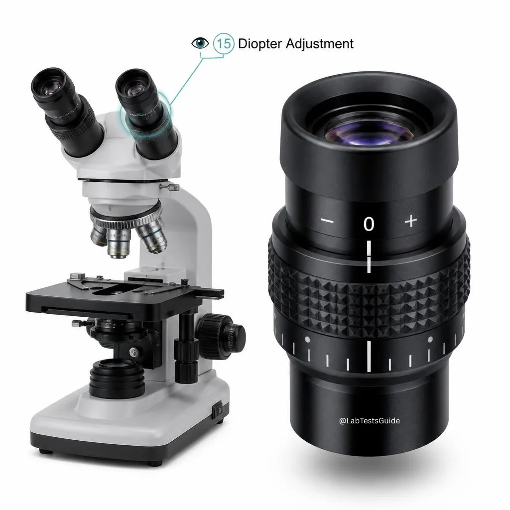
- Eyepiece (ocular): 10x standard, diopter adjustment on binoculars
- Objective lenses: color-coded (4x red, 10x yellow, 40x blue, 100x white oil)
- Coarse/Fine adjustment knobs: coarse for initial focus, fine for sharpening
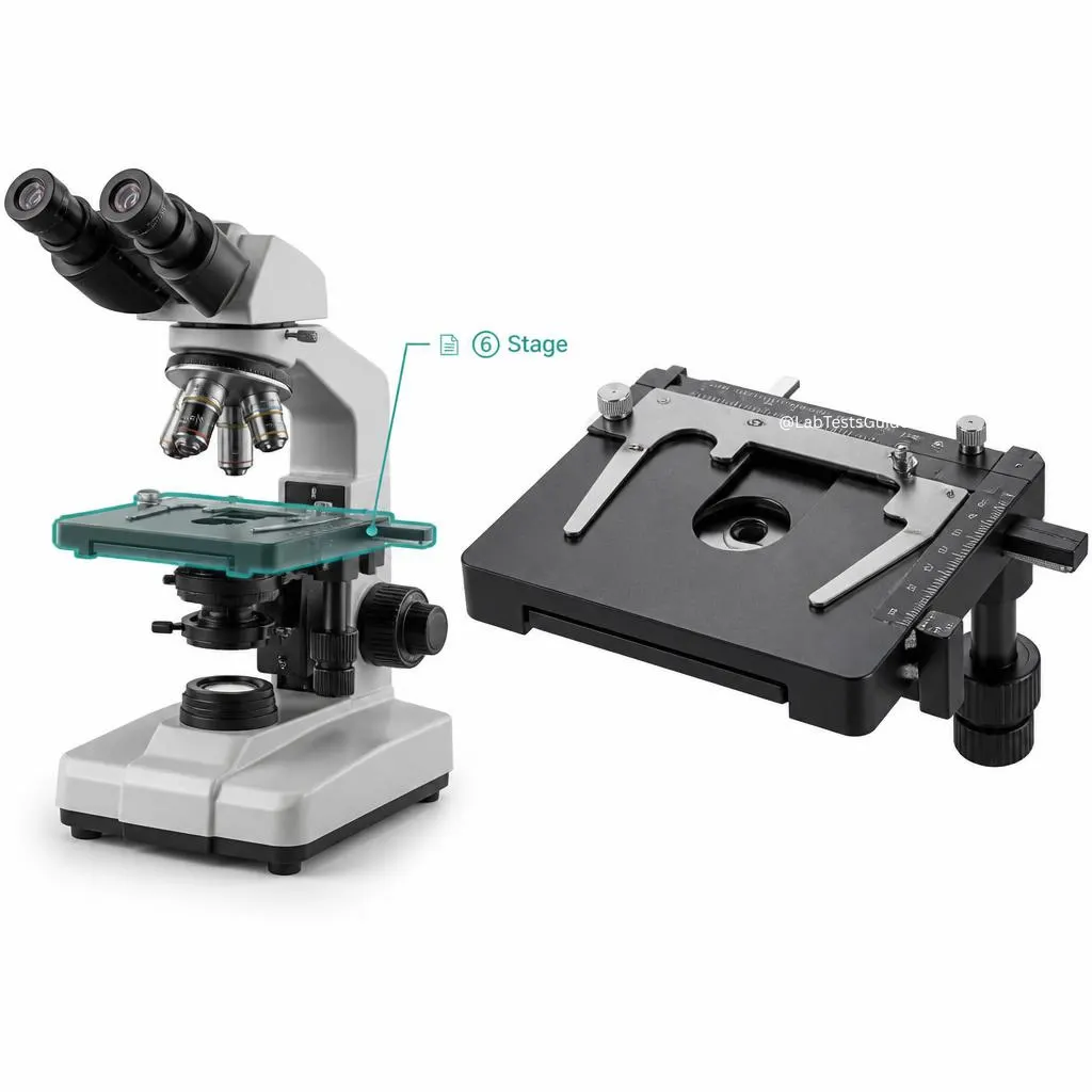
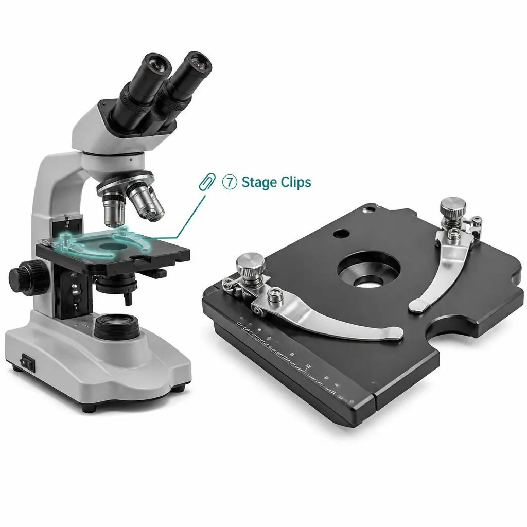
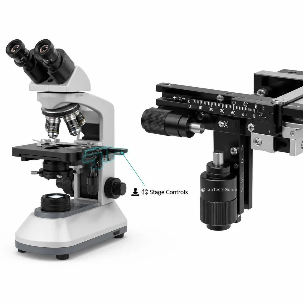
- Stage & stage clips: holds slides; mechanical stage with X-Y knobs
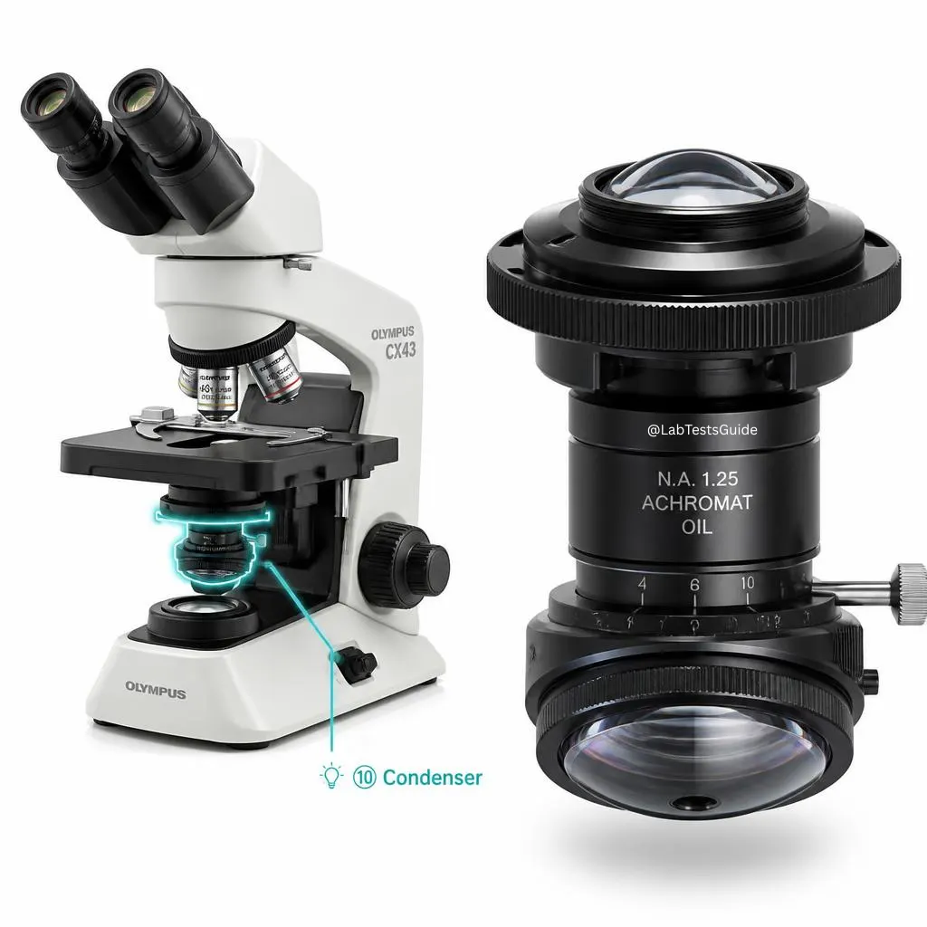
- Condenser & Abbe condenser: focuses light into specimen; NA up to 1.25
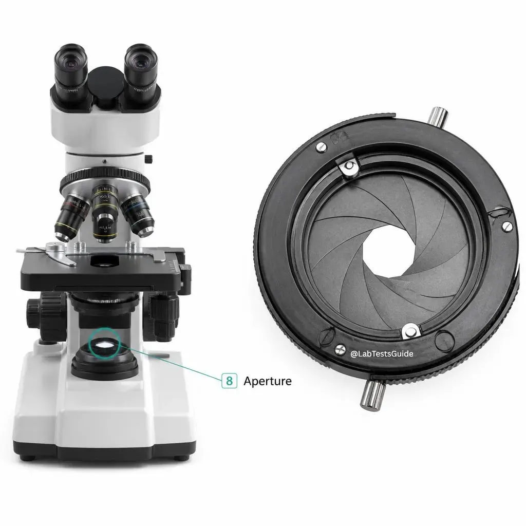
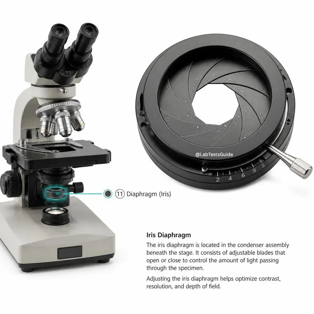
- Iris diaphragm: adjusts light angle and contrast
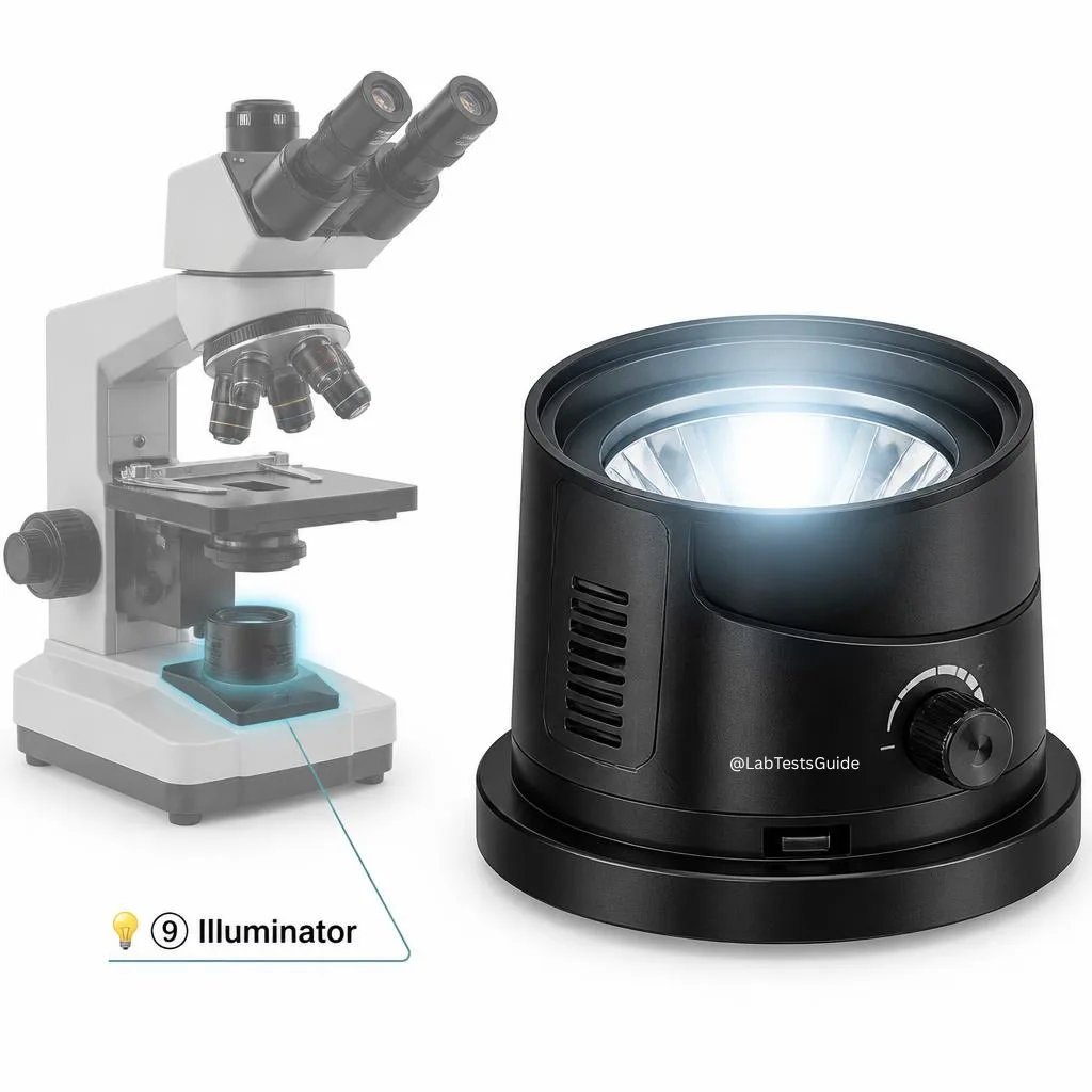
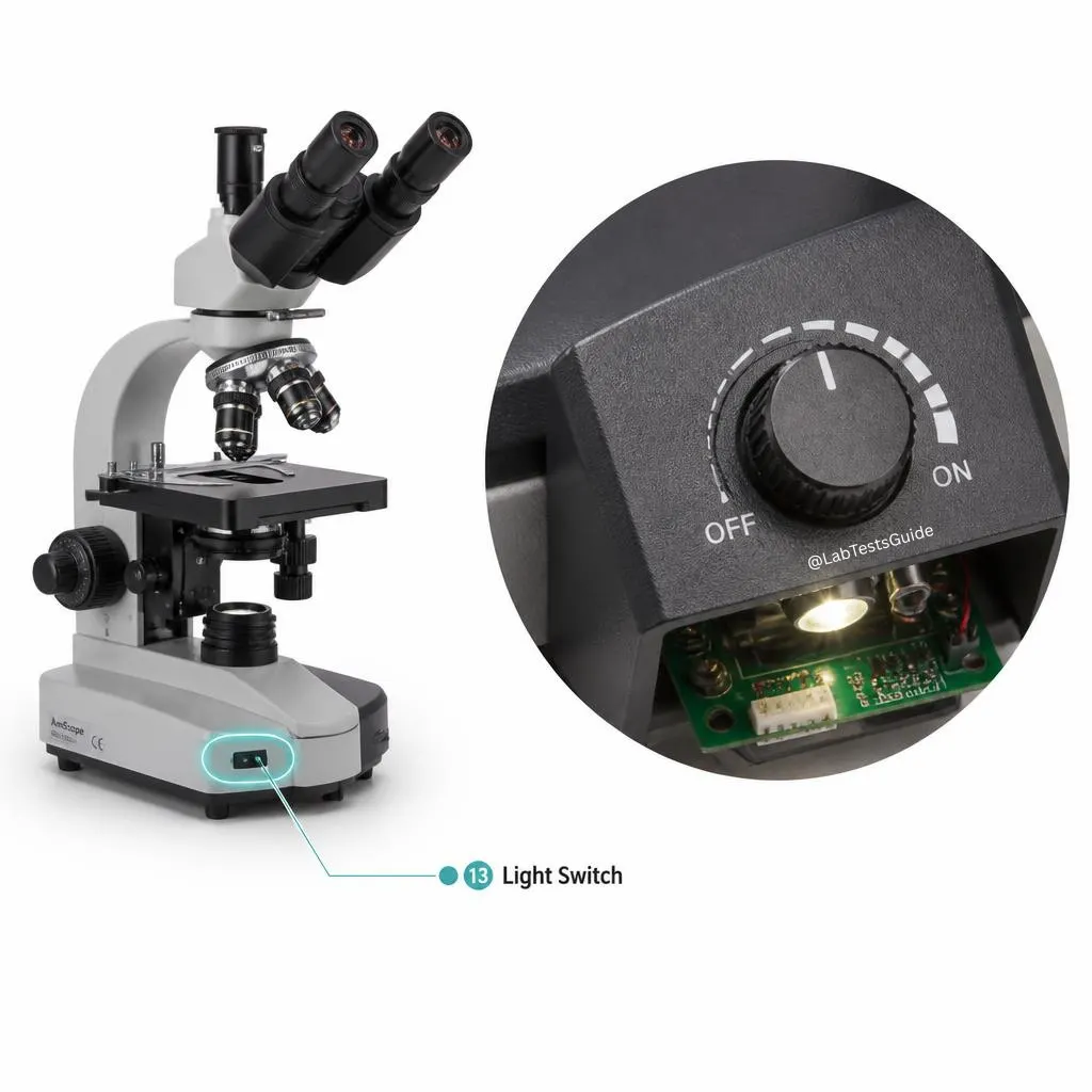
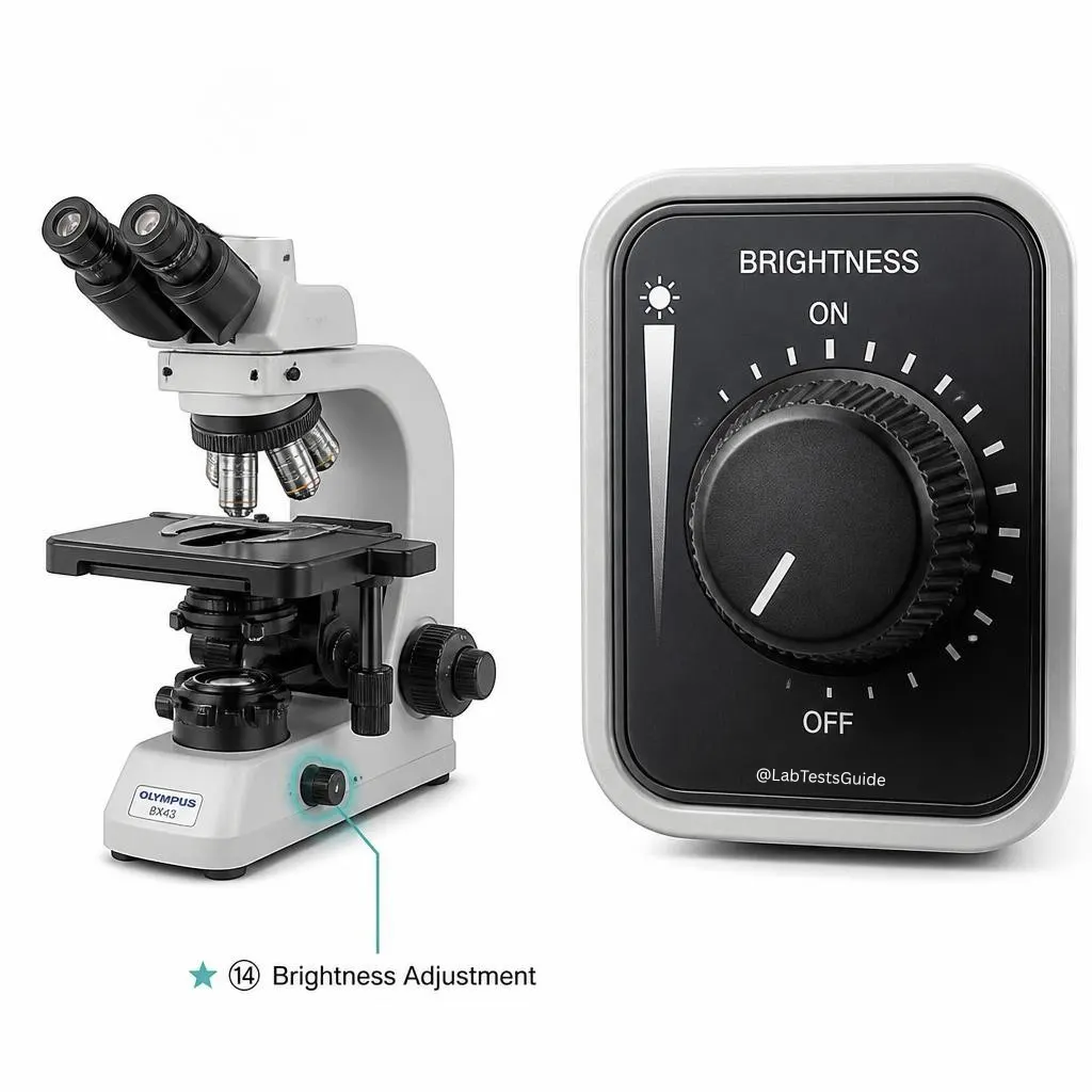
- Illuminator: LED or halogen, brightness control
- Rack stop: prevents slide-objective collision
Download Microscope Diagram PDFs
Optical Principles & Resolution Power
🔬 How the image is formed
Light from illuminator → condenser focuses on specimen → objective lens forms real, inverted, magnified primary image → eyepiece further magnifies to virtual image. Total magnification = Eyepiece × Objective.
Resolution limit (Abbe): d = λ / (2 × NA). Shorter wavelength (blue light, electrons) yields higher resolution. Numerical aperture (NA) = n × sin θ; oil immersion increases NA (n~1.515) → sharper images.
The Diaphragm & Condenser – Mastering Contrast
🎛️ Iris Diaphragm function
Controls the angle and amount of light reaching the specimen. Wide open: brighter but less contrast. Partially closed: increases contrast, depth of field, and reveals fine details. Adjust while viewing — optimal when background is evenly lit without glare.
💎 Condenser (Abbe)
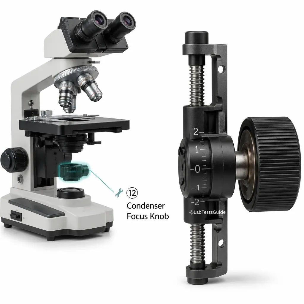
Focuses light into a cone on the specimen. For high power (40x/100x), raise condenser near stage, adjust diaphragm. For low power, lower condenser to match NA. The condenser focus knob fine-tunes light concentration.
Step-by-Step Use & Best Practices
📌 Standard brightfield operation (compound microscope)
- Place on stable surface, turn on illuminator, adjust brightness.
- Rotate nosepiece to lowest power objective (4x).
- Place slide on stage, secure with clips, center specimen over aperture.
- Use coarse focus to raise stage until specimen appears; then fine focus.
- Adjust condenser height & iris diaphragm for optimal contrast.
- Switch to 10x or 40x: only use fine focus knob.
- For 100x oil immersion: place drop of oil, rotate objective into oil, fine focus.
- After use: lower stage, rotate to 4x, clean oil from 100x lens with lens paper, turn off light.
Staining & Specimen Preparation
🎨 Common stains
- Gram stain: Gram+ (purple), Gram- (pink) – bacterial ID
- Acid-fast (Ziehl-Neelsen): Mycobacterium (red)
- H&E: gold standard histology (nuclei blue, cytoplasm pink)
- Methylene blue: simple stain for cell morphology
- Giemsa/Wright: blood smears, malaria, parasites
- DAPI/Fluorescent stains: DNA/protein localization
🧫 Wet mount vs fixed smear
Wet mount: live specimen in water/medium – observe motility. Fixed smear: heat/chemical fixation + staining – permanent slide for detailed structure.
For coverslip: place at 45° angle to avoid air bubbles.
Maintenance, Cleaning & Troubleshooting
🧼 Cleaning protocol
- Lenses: blower → soft brush → lens tissue with 70% ethanol (circular motion).
- Never use paper towels, tissues, or clothing.
- Immersion oil must be removed immediately after 100x use.
- Stage and frame: mild soap solution.
🔧 Common issues & fixes
- Blurry image: clean lenses, adjust diopter, check coverslip thickness (0.17mm).
- Dark field or uneven: raise condenser, center illuminator.
- Stage drifting: tighten tension adjustment collar.
- Black shadow: condenser too low or off-center.
- Cannot focus with 100x oil: check for air bubbles, re-apply oil.
✅ Do's
- Carry with two hands (one on arm, one on base).
- Clean lenses only with specialized Lens Paper.
- Turn off the light when not in use to preserve the bulb.
- Store with a dust cover in a dry environment.
❌ Don'ts
- Never touch lenses with fingers (oils damage coatings).
- Never use the coarse knob on high power (40x or 100x).
- Never leave oil on the 100x lens; clean with Xylene or alcohol after use.
Advanced Microscopy: Super-Resolution & AI
🌟 Super-resolution techniques
STED (20-40 nm), STORM/PALM (10-20 nm), MINFLUX (1-3 nm). Break the Abbe diffraction limit, visualize single molecules and synaptic proteins.
🔬 Cryo-Electron Microscopy (Cryo-EM)
Nobel Prize 2017. Freezes samples in vitreous ice, allows atomic-resolution 3D structures of proteins and viruses without crystallization.
🤖 AI-Powered Digital Microscopy
Deep learning for autofocus, cell segmentation, label-free prediction of stained images, and real-time analysis. Whole-slide scanners & telepathology now standard in 2026 diagnostics.
Real-World Applications Across Fields
🏥 Medical & Clinical
- Pathology: cancer biopsy, tissue diagnosis
- Hematology: blood cell counting, leukemia, malaria
- Microbiology: bacterial ID, fungal analysis
- Reproductive medicine: sperm motility, IVF
🔬 Research & Life Sciences
- Cell biology: organelles, mitosis, cytoskeleton
- Neuroscience: neuron morphology, synapses
- Molecular biology: GFP localization, FISH
🔍 Forensics & Industry
- Trace evidence: hair, fibers, gunshot residue
- Questioned documents: ink, paper fiber analysis
- Semiconductor inspection, metal grain analysis
Frequently Asked Questions (20+ answers)
📚 References & further reading: Murphy D. (2022) Fundamentals of Light Microscopy; Alberts B. Molecular Biology of the Cell; MicrobeNotes.com (Parts, Diaphragm, Types); LabTestsGuide.com; AlmicroMicroscope.com; AlmicroInstruments.com; Nikon MicroscopyU; Zeiss Campus. Updated to 2026 guidelines.
🔬 Conclusion: The microscope remains the most essential tool in life sciences and materials research. From Leeuwenhoek’s single lens to AI-driven super-resolution, mastering its parts, principles, and maintenance unlocks the hidden universe. Whether you are a student, researcher, or clinician, this guide equips you with the knowledge to explore, diagnose, and discover.
© 2026 Microscope Complete Master Guide – Open educational resource. Share, learn, and see the invisible.

9 Comments