Master the Makler Counting Chamber. Learn the 10-micron physics, WHO-compliant SOP protocols, mathematical derivations, calculations, and troubleshooting tips.
Makler Counting Chamber: The Ultimate Guide for Semen Analysis and Sperm Counting
In Andrology and Assisted Reproductive Technology (ART), precision bridges diagnosis and successful patient outcomes. For decades, the Makler Counting Chamber has stood as a cornerstone device for rapid, specialized semen analysis. Unlike deep hematocytometers requiring dilution, the Makler chamber is engineered specifically for assessing undiluted sperm concentration and motility simultaneously. This professional guide maps out its physics, operational protocols, troubleshooting pathways, and clinical metrics.
Chamber Depth
10 µm (0.01 mm)
Fixed, shallow path restricts sperm to a flat horizontal 2D monolayer.
Grid Configuration
1 mm² Area
Subdivided into 100 small squares (0.1 mm x 0.1 mm each).
Sample Aliquot
3 – 10 µL Volume
Requires an ultra-low sample drop size of native, un-diluted semen.
Decimal Math Factor
1 Strip = Millions/mL
Counting 1 row of 10 squares yields the concentration in Millions/mL directly.
What is a Makler Counting Chamber?
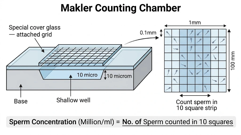
Invented by Dr. Amnon Makler, this reusable slide device eliminates standard hematocytometer inefficiencies. Traditional counting options feature a depth of 100 µm (0.1 mm), permitting spermatozoa to swim out of focus vertically, forcing technicians to kill or chemically immobilize cells prior to tracking. By reducing this depth to 10 µm (0.01 mm), the Makler chamber constrains cells to a single focal plain while maintaining natural motility trajectories.
Why 10 Microns (0.01 mm)?
Monolayer Realization: Human sperm heads possess lengths of ~3.0–5.0 µm and thicknesses of ~1.5–2.5 µm. A 10 µm path is slightly less than a sperm head diameter when swimming vertically but wider than its tail, compelling sperm to lay flat horizontally.
Zero Spatial Overlapping: The compact clearance prevents spermatozoa from swimming over or under one another, ensuring every single cell remains simultaneously visible.
No Focal Blur: Technicians escape the requirement of manipulating fine-focus mechanics continuously during manual tallies, mitigating eye strain and expanding workflow speeds.
History and Architectural Evolution
Prior to the late 1970s, manual reproductive evaluation required tedious 1:20 or 1:200 spermicidal dilutions using formalin or sodium bicarbonate. Dr. Amnon Makler realized that fluid dilution distorted native motility dynamics and added manual errors. Introducing his original prototype validations in Fertility and Sterility (1980), he established a “one-step” framework utilizing four micro-machined corner pillars on a robust frame. While Sefi-Medical Instruments remains the hallmark manufacturer, high-precision laser-serialized clones track across modern lab environments.
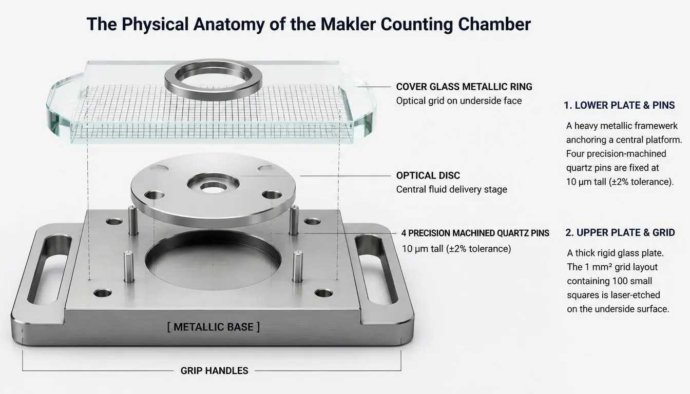
The Physical Anatomy of the Chamber
[ COVER GLASS METALLIC RING ] <– Optical grid on underside face
|
|
v v
+———————————–+
| |
(o) [ OPTICAL DISC ] (o) | <– Central fluid delivery stage
| |
o o | <– 4 Precision Machined Quartz Pins
| |
[ METALLIC BASE ] |
+———————————–+
| |
+——– GRIP HANDLES ———+
Shipped in a protected wooden or padded enclosure, the Makler device consists of five specialized physical components:
1. Lower Plate (Chassis) & Quartz Pins
A heavy metallic framework anchoring a central platform of polished, optically flat glass where the sample drop is loaded. Four precision-machined quartz pins are fixed into the corner boundaries of the base. These pins are exactly 10 µm tall (±2% tolerance) to act as unyielding support pillars for the cover glass.
2. Upper Plate (Cover Glass Ring) & Grid
A thick, rigid glass plate inside a protective metal ring. The 1 mm² grid layout containing 100 small squares is laser-etched on the underside surface of this cover glass. Because the grid contacts the sample directly, both the cells and measurement markers share an identical optical plane under the microscope.
The Chamber Grip Handle: A built-in metal clip extension enabling lab technicians to slide the assembly onto the microscope phase stage without imparting fingerprint smudges or bodily heat transfers, which could cause artificial motility degradation.
The Mathematics of Semen Counting: Geometric Derivations
The core configuration relies on a decimal grid framework, rendering conversion coefficients obsolete during routine clinical counts:
Area of One Small Square: Width (0.1 mm) × Length (0.1 mm) = 0.01 mm².
Volume of One Small Square: Area (0.01 mm²) × Depth (0.01 mm) = 0.0001 mm³ = 10⁻⁴ µL = 10⁻⁷ mL (exactly 1 nanoliter).
Derivation of the 10-Square Linear Strip Rule: Volume of 10 Squares (1 Strip) = 10 × 10⁻⁷ mL = 10⁻⁶ mL = 1/1,000,000 mL.
Because a single row or column equals one-millionth of a milliliter, counting the sperm heads inside 10 continuous squares translates directly into a concentration of Millions per mL (1 cell in 1 strip = 1 × 10⁶ cells/mL).
Derivation of the Full Grid Rule (Oligozoospermia): Volume of All 100 Squares = 100 × 10⁻⁷ mL = 10⁻⁵ mL = 1/100,000 mL.
When dealing with sparse samples, counting all 100 squares and dividing the sum by 10 (or multiplying by 0.1) provides the concentration in Millions/mL. Alternatively, adding five zeros to the raw 100-square total yields cells/mL.
Clinical Makler Diagnostic Suite & Dilution Engine
Compute absolute density metrics, factor custom lab dilutions, and measure total/motile ejaculate yields instantly via unified geometric parameters.
Active Mathematical Formulation Profile
C = [ Raw Tally × (10 ÷ Squares Counted) ] × Dilution Factor (DF)
Arithmetic: [ 25 × 1.00 ] × 1 = 25.00 Million / mL
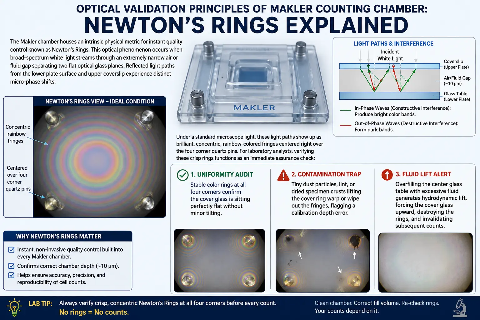
The Makler chamber houses an intrinsic physical metric for instant quality control known as Newton’s Rings. This optical phenomenon occurs when broad-spectrum white light streams through an extremely narrow air or fluid gap separating two flat optical glass planes. Reflected light paths from the lower plate surface and upper coverslip experience distinct micro-phase shifts:
In-Phase Waves (Constructive Interference): Produce bright color bands.
Out-of-Phase Waves (Destructive Interference): Form dark bands.
Under a standard microscope light, these light paths show up as brilliant, concentric, rainbow-colored fringes centered right over the four corner quartz pins. For laboratory analysts, verifying these crisp rings functions as an immediate assurance check:
Uniformity Audit: Stable color rings at all four corners confirm the cover glass is sitting perfectly flat without minor tilting.
Contamination Trap: Tiny dust particles, lint, or dried specimen crusts lifting the cover ring warp or wipe out the fringes, flagging a calibration depth error.
Fluid Lift Alert: Overfilling the center glass table with excessive fluid generates hydrodynamic lift, forcing the cover glass upward, destroying the rings, and invalidating subsequent counts.
Standard Operating Procedure (SOP): Step-by-Step Execution
To secure reproducible data matching modern global baseline expectations, technicians must follow this checklist[cite: 1]:
Phase 1: Pre-Analytical Preparation
1. Liquefaction: Allow raw ejaculate to completely liquefy in an incubator (20–30 minutes at 37°C) to break down mucosal strings.
2. Homogenization: Swirl the specimen vial gently using smooth circular loops. Avoid aggressive vortexing, which shears delicate sperm flagella and induces air bubble pockets.
3. Thermal Shielding: Pre-warm the metallic base plate and cover glass at 37°C for 5–10 minutes on a warming table. Cold glass prompts “cold shock,” severely arresting motility metrics.
Phase 2: Pipetting & Chamber Loading
1. Aliquot Delivery: Use a digital micropipette to draw a 3–10 µL drop from the core of the sample and deposit it directly in the center of the optical glass disc.
2. Immediate Sealing: Pick up the cover glass ring by its outer border and place it straight down onto the corner quartz pins without hesitation.
CRITICAL TIME GATEWAY (The 5-Second Warning): Any delay between depositing the fluid sample and seating the upper glass cover generates convective heat currents. Active sperm rapidly collect and pack directly inside the central viewing focus. Pausing for 30 seconds can falsely elevate concentration variables by 107% and motility values by 42%. The glass must be dropped securely within 0–5 seconds.
3. Lock Verification: Apply gentle pressure at the perimeter ring coordinates until concentric rainbow-colored Newton’s Rings resolve clearly at the corner pins, ensuring an exact 10 µm sample thickness.
Phase 3: Microscopic Alignment & Tallying
1. Focus Setup: Place the chamber on the stage. Engage a 20x objective lens (yielding 200x total magnification via a 10x eyepiece), which balances field width with tracking resolution. Phase-contrast microscopy is recommended to illuminate transparent sperm heads without staining. If phase contrast is unavailable, constrict the field diaphragm to enhance image contrast manually. Allow 1 minute for thermal re-stabilization on a heated stage.
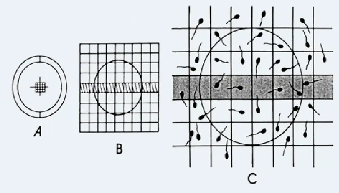
2. Standard Linear Count: Count all intact sperm heads across one horizontal strip of 10 squares. That value equals the global concentration in Millions/mL directly. Repeat the assay on a separate independent strip and average the results to verify consistency.
3. Polyzoospermic Modification (Extreme Density): In highly crowded fields, count sperm within only 5 small squares and multiply the final sum by 2 to ascertain the Millions/mL concentration value. For extreme cases, dilute a separate aliquot 1:10 with saline and multiply the final chamber count by 10.
4. Border Line Boundary Rule (Left/Upper Inclusion Grid Rule): To prevent double-counting or omitting cells resting on grid borders, count sperm whose heads lie completely inside a square, or rest directly on the top or left border lines. Disregard sperm heads touching the bottom or right lines. Count only intact spermatozoa with tails attached; ignore loose tails or detached pinheads during primary tallying.
Measuring and Classifying Sperm Motility
The 10 µm boundary restricts vertical velocity vectoring, making it easier to monitor individual sperm pathways across four standard WHO kinetic categories[cite: 1]:
Category A (Rapid Progressive): Sperm cells traveling dynamically forward in straight linear headings or wide sweeping circles, displaying speeds exceeding 25 µm/s at 37°C[cite: 1].
Category B (Slow Progressive): Sperm cells traveling forward, but with reduced velocity or along curved, meandering paths[cite: 1].
Category C (Non-Progressive): Active tail beating is present, but lacks forward progression (e.g., swimming in tight localized loops or vibrating in place)[cite: 1].
Category D (Immotile): Completely still; displays zero active flagellar movement[cite: 1].
Motility Protocol: Because kinetic movement declines post-ejaculation, analyze motility immediately after loading[cite: 1]. Select a square or strip and utilize a two-step tally: count all rapid and slow progressive cells first, then tally the stationary immotile heads in that same field. Count at least 200 sperm across separate grid coordinates to maintain accuracy[cite: 1].
The Ségre-Silberberg (Segregation) Effect: When fluid suspensions pass into an ultra-narrow 10 µm boundary, axial hydrodynamic forces can cause particles to distribute unevenly. Sperm heads tend to gather toward the central axis of the moving fluid column rather than along the peripheral boundaries, risking minor overestimation biases if the sample drop spreads unevenly. Viscosity Restrictions: Hyper-viscous or poorly liquefied semen samples struggle to spread uniformly across a narrow 10 µm gap. Cellular debris can clump near the entry margin, filtering out sperm cells and skewing downstream counts.
Integration with Computer-Assisted Semen Analysis (CASA) Systems
Modern fertility labs link manual chambers with automated CASA platforms combining digital video capture and edge-detection algorithms. The Makler profile acts as a robust hardware interface for automation:
Focal Tracking Lock: Because the 10 µm clearance prevents vertical tracking loss, sperm rarely swim out of focus, allowing software to maintain continuous track locks on individual cells.
Enhanced Image Contrast: Clear glass layers produce bright halos around sperm heads under phase-contrast optics, simplifying automated tracking.
Kinematic Calibration Constants: Software modules can establish repeatable environments for calculating kinematic variables like Curvilinear Velocity (VCL), Straight-Line Velocity (VSL), and Average Path Velocity (VAP). Capture frequencies must be set to 30–60 Hz to document lateral head displacement without motion blur. Turn off digital grid overlays when utilizing physical etched lines to calibrate pixel-to-micron camera scaling.
Meticulous Cleaning, Decontamination, and Maintenance
To preserve calibration accuracy and eliminate carryover patient contamination, observe this workflow:
Mechanical Wipe: Use the specialized soft brush provided with the kit, dipped in distilled water or an approved non-corrosive, aldehyde-free disinfectant. Gently clear the central glass base and the etched coverslip grid. Sponge off residual droplets with a squeezed, lint-free swab.
Optical Surface Polish: Gently rub the elements in circular loops using premium lens tissue (e.g., Kimwipes) to dissolve oils. Standard paper tissues leave micro-scratches. Never touch the quartz pins, as abrasive degradation alters the depth profile permanently.
Pathogen Decontamination: If exposed to infectious biohazards (HIV, Hepatitis, HSV), submerge the separated glass items in 70% Isopropyl Alcohol or Ethanol for 10 minutes. Avoid chlorine bleach (sodium hypochlorite), which corrodes the frame assembly and degrades pin adhesives. The chamber cannot withstand steam autoclaving or high-pressure sterilization cycles. Use low-temperature ethylene oxide (EtO) gas if absolute sterilization is required.
Quality Control, Calibration Verification, and Validation Kits
To preserve laboratory accreditation (such as CLIA, CAP, or ISO 15189), quantitative equipment must undergo validation audits:
Certified Control Beads: Technicians can verify volumetric performance using certified polymer microsphere suspensions (e.g., QwikCheck Beads) calibrated to known density targets mimicking sperm head parameters.
Validation Workflow: Roll the control vial smoothly between hands to distribute beads evenly without generating bubble pockets. Pipette 3.5–5 µL into the clean chamber, verify corner Newton’s Rings, and count the spheres across 10 squares under 200x magnification. Calculated bead counts must tightly match the target verification guidelines: Target Concentration = Mean Assay Reference Value (± Acceptable Standard Deviation Limit)
If results drift outside the target range, check for residue buildup on the corner pins or contact an authorized service center for professional recalibration.
Clinical Interpretations, Reference Values, and Case Studies
Interpret results using the standard reference thresholds from the 6th Edition of the WHO Semen Processing Manual[cite: 1]:
Semen Metric
WHO 6th Edition Lower Reference Thresholds
Sperm Concentration
≥ 15 × 10⁶ spermatozoa per mL[cite: 1]
Total Motility (PR + NP)
≥ 40%[cite: 1]
Progressive Motility (PR)
≥ 32%[cite: 1]
Total Tally per Ejaculate
≥ 39 × 10⁶ sperm cells per total volume
Veterinary Applications in Animal Husbandry
While optimized for human care, the Makler chamber is widely utilized across commercial livestock breeding and animal clinics:
Equine Stud Management: Stallions yield high ejaculate volumes (50–150 mL) with variable cell densities. The Makler design allows on-site veterinary technicians to calculate concentrations within minutes of collection, helping optimize fresh semen transport dilution ratios.
Bovine Insemination Centers: Commercial cattle facilities handle frozen straws stored in liquid nitrogen. Upon thawing, technicians leverage pre-warmed Makler slides to perform rapid viability checks, ensuring straws meet strict progressive motility standards prior to field insemination.
Canine Reproductive Focus: Specialized veterinary teams use the device to calibrate chilling and shipping volumes for valuable stud dogs. Rapid single-plane tracking minimizes the time sensitive canine sperm spends outside temperature-controlled environments.
Frequently Asked Questions (FAQ)
Q1: What is the Makler chamber?+
A: The Makler chamber is a disposable plastic device used in reproductive medicine to evaluate the concentration and motility of sperm in semen samples.
Q2: How does the Makler chamber work?+
A: The chamber consists of two chambers separated by a coverslip, with the upper chamber having a depth of 10 microns and the lower chamber having a depth of 20 microns. A semen sample is placed on the coverslip, which allows the sperm to settle into the chambers by capillary action. The sperm in the chambers are then counted and assessed under a microscope.
Q3: Who invented the Makler chamber?+
A: The Makler chamber was invented by Dr. Michael Makler, an Israeli physician, in the early 1980s.
Q4: Why is the Makler chamber preferred over other types of counting chambers?+
A: The Makler chamber allows for a more accurate assessment of sperm concentration and motility compared to other types of counting chambers. It is also easy to use, disposable, and cost-effective.
Q5: How long can the Makler chamber be stored?+
A: Indefinitely. Because it is made of durable optical glass and metal rather than perishable parts, its shelf life is virtually unlimited as long as it is kept clean, dry, scratch-free, and stored in its protective padded case.
Q6: How is the Makler chamber disposed of after use?+
A: The Makler chamber is not disposable; it is reusable.
Q7: What are the advantages of using the Makler chamber in evaluating male infertility?+
A: The Makler chamber allows for an accurate and reliable assessment of sperm concentration and motility, which are important factors in evaluating male infertility. It is also easy to use and cost-effective.
Q8: Are there any potential limitations or sources of error when using the Makler chamber?+
A: Like all diagnostic tests, there is a potential for error when using the Makler chamber. Possible sources of error include inadequate sample collection or preparation, technical errors during the assessment process, and variation in results due to biological factors such as sperm morphology.
Q9: How long does it take to perform a Makler chamber evaluation?+
A: The Makler chamber evaluation typically takes about 10-15 minutes to complete.
Q10: Is the Makler chamber available worldwide?+
A: Yes, the Makler chamber is widely available in reproductive clinics and laboratories around the world.
Q11: Are there any special considerations or precautions that should be taken when using the Makler chamber?+
A: It is important to follow the instructions for use provided with the Makler chamber to ensure accurate and reliable results. The device should be used only by trained personnel in a laboratory setting, and appropriate safety measures should be taken when handling semen samples.
Q12: Can the Makler chamber be used to evaluate sperm in frozen semen samples?+
A: Yes, the Makler chamber can be used to evaluate sperm in fresh or frozen semen samples.
Q13: Can the Makler chamber be used to evaluate sperm from other species besides humans?+
A: Yes, the Makler chamber can be used to evaluate sperm from other species, including animals. However, the chamber should be selected based on the appropriate dimensions for the sperm being evaluated.
Q14: Can I use standard lab coverslips with the Makler chamber base?+
A: No. Standard coverslips lack the precision weight, optical flatness, and direct etched grids needed for this system. The specialized Makler cover glass is calibrated to rest level across the quartz pins without bending. A standard coverslip alters the clearance gap, compromising accuracy.
Q15: Why do sperm exhibit uniform streaming paths across the grid?+
A: Streaming patterns indicate chamber overfilling (exceeding 5–10 µL), which creates hydrodynamic pressure that causes the fluid layer to drift. It can also occur if the cover glass isn’t sitting flat on the quartz pins. Clean the plates completely and reload using a smaller 3–4 µL drop.
Q16: How frequently must a Makler chamber undergo recalibration?+
A: For accredited environments (ISO 15189 or CAP), perform a calibration check annually. If the chamber suffers a physical drop, shows visual wear, or fails weekly control bead audits, remove it from service immediately for professional servicing.
Q17: Is the system effective for examining retrograde ejaculation samples?+
A: Yes. The Makler chamber is clinically utilized to check urine samples for the presence of sperm cells in cases of suspected retrograde ejaculation.
Q18: What are the primary purchasing metrics for identifying genuine chambers?+
A: Look for distinct laser-etched grids that resist alcohol cleaning, unique laser-etched manufacturing serial numbers for laboratory tracking, and a specific heft that ensures stability under the microscope. Authentic units generally retail between $600 to $900 USD.
The website covers a wide range of lab tests, including blood tests, urine tests, stool tests, and imaging tests such as X-rays and CT scans. It also provides information about different health conditions and diseases, as well as tips for maintaining good health.
It's important to note that while labtestsguide.com may provide valuable information about lab tests and their interpretation, it's always best to consult with a healthcare professional if you have any concerns or questions about your lab results. We can provide personalized guidance and advice based on your individual health status and medical history.



One Comment