CSF Analysis Test Purpose, Procedure, Result Interpretation, Reasult Meaning and Report Format
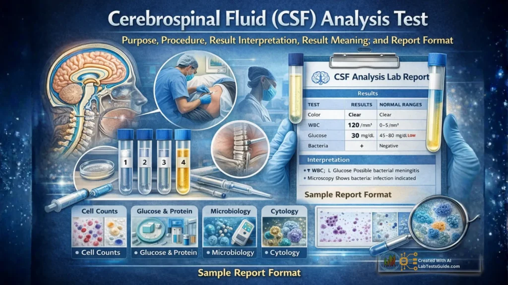
A cerebrospinal fluid (CSF) test analyzes the clear fluid that surrounds the brain and spinal cord to diagnose central nervous system (CNS) problems, such as infections (meningitis), inflammation (MS), bleeding, or cancer, by looking for abnormal cells, proteins, sugar, bacteria, or pressure changes, often collected through a lumbar puncture.
CSF Analysis Test Quick Facts:
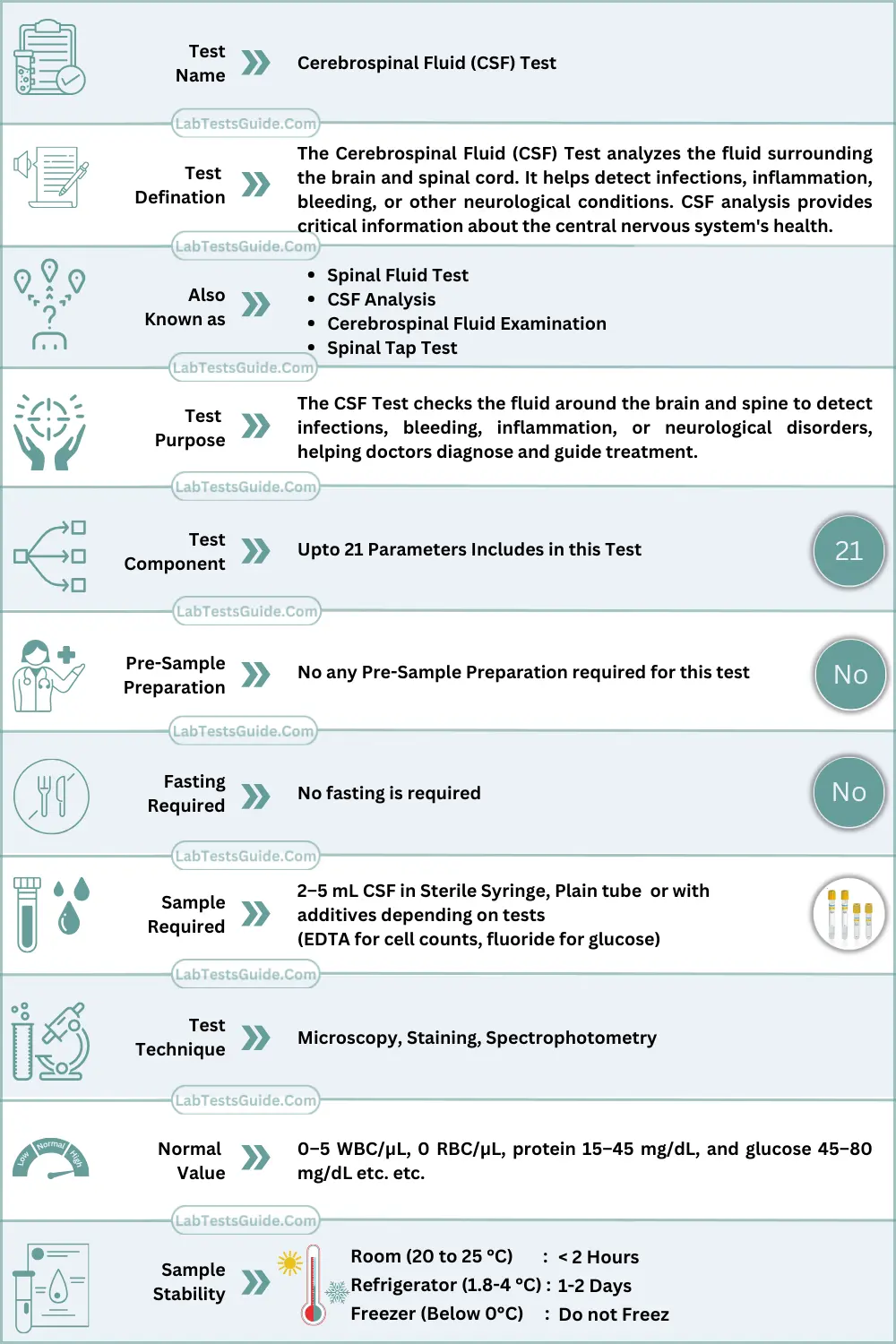
What is Cerebrospinal Fluid (CSF) Analysis Test?
The Cerebrospinal Fluid (CSF) Test analyzes the fluid surrounding the brain and spinal cord. It helps detect infections, inflammation, bleeding, or other neurological conditions. CSF analysis provides critical information about the central nervous system’s health.
Why is Cerebrospinal Fluid (CSF) Analysis Test Done?
For Patients / General Use
- Persistent headache or severe headache
- Fever with neck stiffness
- Nausea and vomiting with neurological symptoms
- Confusion, seizures, or altered consciousness
- Symptoms of multiple sclerosis (e.g., numbness, weakness)
- Suspected bleeding in the brain or spinal cord
For Doctors / Clinical Use
- Diagnose meningitis, encephalitis, or CNS infections
- Detect subarachnoid hemorrhage or intracranial bleeding
- Monitor autoimmune or inflammatory neurological disorders
- Evaluate malignancies affecting the CNS (leukemia, lymphoma, metastatic cancer)
- Guide therapeutic decisions and monitor response to treatment
How the Cerebrospinal Fluid (CSF) Test Works (Principle / Methodology)
CSF is collected via lumbar puncture and analyzed for:
- Cell counts and differential: Microscopy using automated counters or manual methods
- Protein & glucose levels: Colorimetric assays or enzymatic methods
- Microbiological evaluation: Culture, Gram stain, PCR, and antigen detection
- Immunological markers: ELISA for antibodies or oligoclonal bands
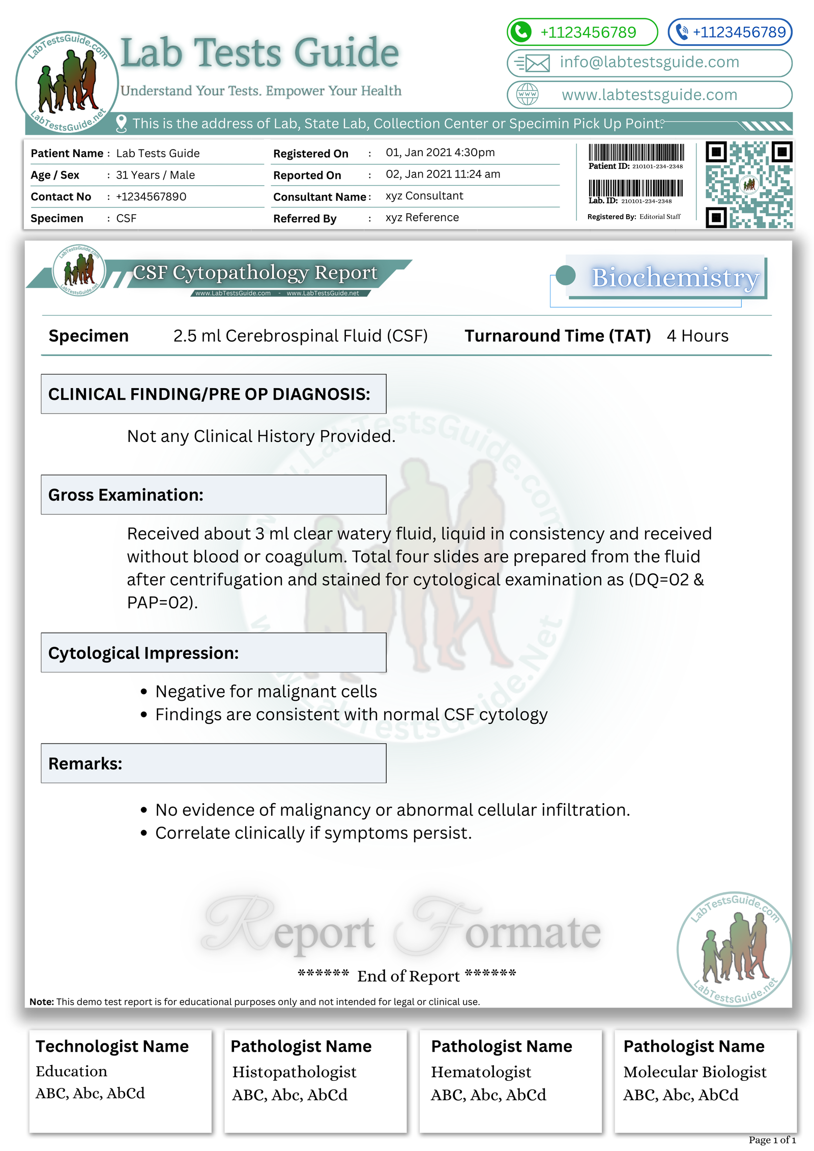
- Cytology: Detect malignant cells
The combination of biochemical, cytological, and microbiological analysis provides a complete picture of CNS health.
Cerebrospinal Fluid (CSF) Test Specimen Requirements & Collection
- Specimen type: Cerebrospinal fluid
- Tube type: Sterile, labeled tubes (plain, EDTA, or fluoride depending on tests)
- Volume: 2–5 mL
- Patient preparation: Explain procedure, position in lateral decubitus, no fasting required
- Collection steps:
- Patient positioned safely
- Lumbar puncture performed under sterile conditions
- Collect CSF in sequential tubes (Tube 1: chemistry; Tube 2: microbiology; Tube 3: cell count)
- Transport & storage:
- Transport immediately at room temperature for cell counts
- Refrigerate biochemical specimens if delay >1 hour
- Avoid shaking
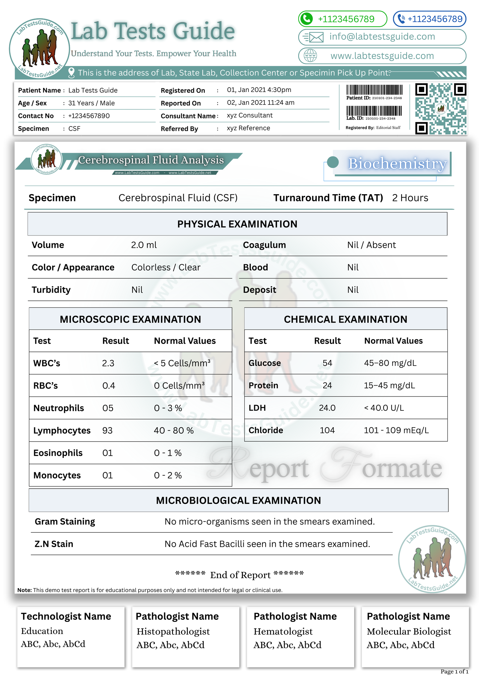
Cerebrospinal Fluid (CSF) Test Reference Ranges
1. Physical Examination
| Parameter | Normal Result / Value |
|---|---|
| Color | Clear, colorless |
| Clarity / Appearance | Transparent / Clear |
| Odor | None / Odorless |
| Volume | 1–2 mL (typically collected 2–5 mL) |
| Pressure (Opening) | 70–180 mm H₂O (adults) |
2. Chemical Examination
| Parameter | Normal Value |
|---|---|
| Glucose | 45–80 mg/dL (or 2.5–4.4 mmol/L) — ~60–70% of blood glucose |
| Protein (Total) | 15–45 mg/dL (0.15–0.45 g/L) |
| LDH | 10–22 mg/dL (0.9–2.5 mmol/L) |
| Chloride | 118–132 mEq/L |
| Osmolality | 280–300 mOsm/kg |
3. Microscopic Examination
| Parameter | Normal Result |
|---|---|
| Red Blood Cells (RBCs) | 0 / mm³ |
| White Blood Cells (WBCs) | 0–5 / mm³ (mostly lymphocytes) |
| Cell Type | Mostly lymphocytes; occasional monocytes |
| Cytology | No abnormal / malignant cells |
| Crystals | None |
4. Microbiological Examination
| Parameter | Normal Result |
|---|---|
| Gram Stain | No bacteria seen |
| Culture | No growth (sterile) |
| PCR / Antigen Tests | Negative for viruses/bacteria/fungi |
| India Ink (for Cryptococcus) | Negative |
Cerebrospinal Fluid (CSF) Test Interpretation of Results
High Levels (Causes & Clinical Significance)
- Protein: Infection, inflammation, demyelination, tumor
- WBC: Bacterial or viral meningitis, autoimmune CNS disease
- RBC: Subarachnoid hemorrhage, traumatic tap
Differential diagnoses: Bacterial meningitis, multiple sclerosis, CNS malignancy, intracranial bleeding
Clinical relevance: Helps confirm infection, bleeding, or inflammatory process
Low Levels (Causes & Clinical Significance)
- Protein: CSF leak, overhydration
- Glucose: Hypoglycorrhachia seen in bacterial meningitis or malignancy
Differential diagnoses: CSF leak, severe systemic hypoglycemia
Clinical relevance: Guides urgent interventions or further diagnostic imaging
CSF Analysis Test Errors
1. Pre-Analytical Errors
These occur before the sample reaches the lab and are the most common source of errors.
| Error Type | Cause / Example | Impact on Result |
|---|---|---|
| Improper collection | Traumatic lumbar puncture, too little CSF | Blood contamination, inaccurate cell counts, false protein increase |
| Incorrect tube use | Mixing tubes for chemistry, microbiology, and cytology incorrectly | Cross-contamination, wrong test results |
| Delayed transport | CSF left at room temperature for long periods | Cell lysis, decreased glucose, false low WBC count |
| Wrong labeling / misidentification | Patient mislabeling or tube mix-up | Misdiagnosis or reporting error |
| Insufficient volume | Less than 1–2 mL collected per tube | Some tests may not be performed, repeat procedure required |
| Exposure to air / light | Sample left uncovered or in light | May affect certain chemical tests like glucose |
| Temperature errors | Not transported on ice for special tests | Degradation of glucose or labile proteins |
2. Analytical Errors
These occur during actual laboratory testing.
| Error Type | Cause / Example | Impact on Result |
|---|---|---|
| Instrument calibration errors | Poorly maintained analyzers | Incorrect protein, glucose, or cell count |
| Reagent issues | Expired or contaminated reagents | False chemical values |
| Incorrect dilution | Manual pipetting errors | WBC/RBC counts inaccurate |
| Timing errors | Delayed centrifugation or staining | Cell lysis, inaccurate microscopy results |
| Operator errors | Miscounting cells, misinterpreting Gram stain | False positive/negative results |
| Interference | Blood contamination, high protein, or xanthochromia | Alters chemical measurements |
3. Post-Analytical Errors
These occur after the test has been performed, mostly in reporting or communication.
| Error Type | Cause / Example | Impact on Result |
|---|---|---|
| Transcription errors | Mistyped values in report | Wrong patient results delivered |
| Delayed reporting | Lab backlog or communication delays | Treatment may be delayed |
| Misinterpretation | Clinician misreads values or reference ranges | Misdiagnosis |
| Loss of results | Electronic or paper record errors | Repeat sampling may be needed |
| Wrong reference ranges used | Pediatric vs adult, lab-specific variations | Confusing normal vs abnormal results |
Interfering Factors
- Hemolysis: Falsely increases RBC count and protein
- Lipemia: Rare; may interfere with colorimetric glucose assays
- Icterus: Minimal effect
- Medications: Antimicrobials may suppress culture growth
- Sample handling: Delay >1 hr can alter cell counts
- Biological variations: Age, pregnancy, or recent CNS trauma
Cerebrospinal Fluid (CSF) Test Critical Values / PANIC Values
- Glucose <30 mg/dL
- Protein >500 mg/dL
- WBC >1000/µL
- RBC >1000/µL (non-traumatic)
Follow institution policy for urgent reporting.
🧠 AI-Powered Test Result Analysis:
Understand your Cerebrospinal Fluid (CSF) Analysis Test Results
AI-powered Lab Test Results Meaning tool 🤖
📥 Download Cerebrospinal Fluid (CSF) Analysis Test Lab Report Format
Get the demo report format for Cerebrospinal Fluid (CSF) Analysis Test in your preferred format. These templates are fully editable and professional.
How to Download?
| File Description | Actions |
|---|---|
Cerebrospinal Fluid (CSF) Analysis Test Report Format (Image) | |
Cerebrospinal Fluid (CSF) Analysis Test Report Format (MS Word) | |
Cerebrospinal Fluid (CSF) Analysis Test Report Format (MS Excel) | |
Cerebrospinal Fluid (CSF) Analysis Test Report Format (PDF) | |
ABC File Download | |
Purchase ABC Book |
| File Description | Actions |
|---|---|
Cerebrospinal Fluid (CSF) Cytology Test Report Format (Image) | |
Cerebrospinal Fluid (CSF) Cytology Test Report Format (MS Word) | |
Cerebrospinal Fluid (CSF) Cytology Test Report Format (MS Excel) | |
Cerebrospinal Fluid (CSF) Cytology Test Report Format (PDF) | |
ABC File Download | |
Purchase ABC Book |
| File Description | Actions |
|---|---|
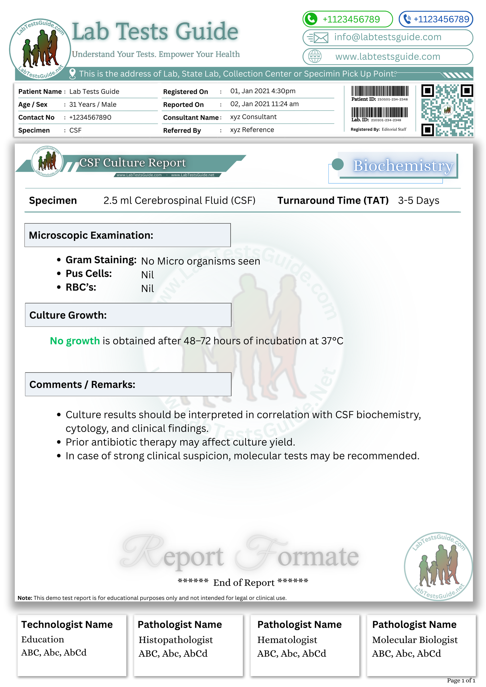
Cerebrospinal Fluid (CSF) Culture No Growth Report Format (Image) | |
Cerebrospinal Fluid (CSF) Culture No Growth Report Format (MS Word) | |
Cerebrospinal Fluid (CSF) Culture Nor Growth Report Format (MS Excel) | |
Cerebrospinal Fluid (CSF) Culture Nor Growth Report Format (PDF) | |
ABC File Download | |
Purchase ABC Book |
| File Description | Actions |
|---|---|
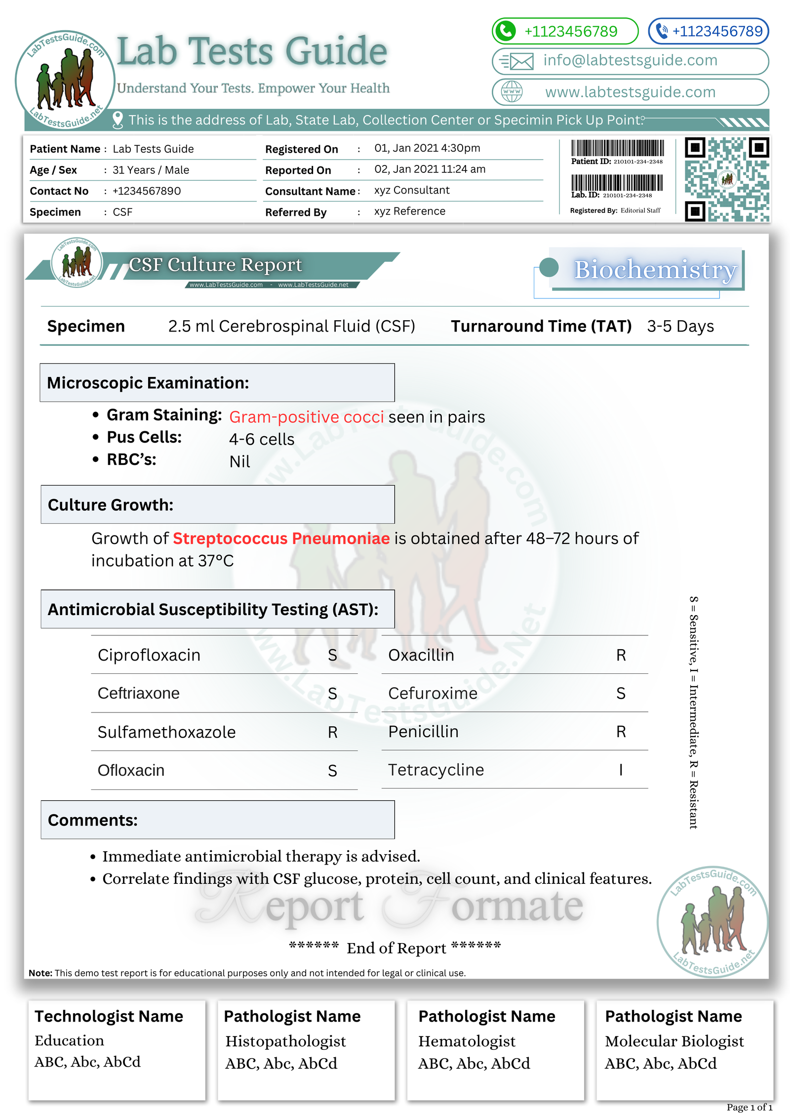
Cerebrospinal Fluid (CSF) Culture Test Positive Growth Report Format (Image) | |
Cerebrospinal Fluid (CSF) Culture Test Positive Growth Report Format (MS Word) | |
Cerebrospinal Fluid (CSF) Culture Test Positive Growth Report Format (MS Excel) | |
Cerebrospinal Fluid (CSF) Culture Test Positive Growth Report Format (PDF) | |
ABC File Download | |
Purchase ABC Book |
Cerebrospinal Fluid (CSF) Test Nursing / Phlebotomy Notes
- Use sterile tubes; label immediately
- Collect 3–5 mL CSF in sequential tubes
- Transport promptly; maintain proper temperature
- Avoid shaking or contamination
- Monitor patient for post-lumbar puncture headache or bleeding
Cerebrospinal Fluid (CSF) Test Lab Student Key Points
- CSF is obtained via lumbar puncture
- Analyze color, clarity, cells, protein, glucose, microbiology
- Reference ranges differ by age and lab
- Rapid transport is essential to maintain cell integrity
- Elevated WBC suggests infection, RBC suggests bleeding
Does CSF collection hurt?
Mild discomfort; local anesthesia is used.
Do I need to fast?
No fasting is required.
How long for results?
Typically 1–3 days.
Can medication affect the test?
Yes, antibiotics can reduce bacterial detection.
Is a lumbar puncture safe?
Generally safe; complications are rare.
Why is CSF glucose low in infections?
Bacteria consume glucose, lowering levels.
Can CSF test detect cancer?
Yes, malignant cells may be found in cytology.
How can I download CSF Test Report templates?
Select your preferred format (PDF, Word, Excel, or PNG) and click the download button. You can also preview the files before downloading.
Are the lab report formats editable?
Yes, our MS Word and MS Excel templates are fully editable, allowing you to add your own lab logo and details.