X Ray, also known as X-radiation, are a form of electromagnetic radiation. They have shorter wavelengths and higher energy levels than visible light. X-rays were discovered by Wilhelm Conrad Roentgen in 1895, and they have since found numerous applications in medicine, industry, and scientific research.
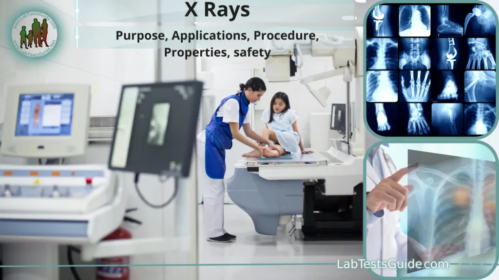
- X-rays are a form of electromagnetic radiation, with shorter wavelengths than visible light.
- They were discovered by Wilhelm Conrad Roentgen in 1895, earning him the first Nobel Prize in Physics.
- X-rays have a wide range of applications, including medicine, industry, and scientific research.
- X-ray machines consist of an X-ray tube that produces the radiation and a detector to capture the X-ray images.
- The ability of X-rays to penetrate matter makes them valuable for imaging the interior of objects and organisms.
- Medical X-ray imaging is used for diagnosing conditions like fractures, infections, and tumors.
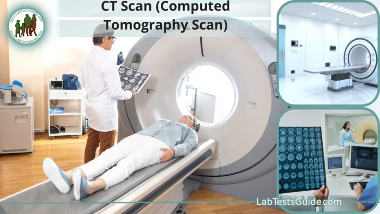
- Computed Tomography (CT) scans use X-rays to create detailed cross-sectional images of the body.
- Fluoroscopy is a real-time X-ray imaging technique used for procedures like angiography and barium studies.
- X-rays are used in radiation therapy to treat cancer by targeting and damaging cancerous cells.
- In industry, X-rays are employed for non-destructive testing of materials and welds.
- They are also used for quality control in manufacturing processes.
- X-ray scanners at airports are used to inspect baggage for security purposes.
- X-ray crystallography is a scientific technique that uses X-rays to determine the atomic and molecular structure of crystals.
- X-rays are part of the electromagnetic spectrum, which includes gamma rays, ultraviolet rays, visible light, and more.
- The energy of X-rays can be adjusted to suit specific applications, from low-energy dental X-rays to high-energy radiation therapy.
- Radiographers and radiologic technologists are trained professionals who operate X-ray equipment and ensure patient safety.
- Lead aprons and other shielding materials are used to protect healthcare workers and patients from unnecessary radiation exposure.
- Radiation doses in X-ray procedures are carefully controlled to minimize health risks.
- Digital X-ray imaging has largely replaced traditional film-based X-rays, offering faster results and lower radiation doses.
- Three-dimensional (3D) X-ray imaging techniques, like cone-beam CT, provide detailed volumetric images.
- X-rays are used in archeology and paleontology to examine the interior of artifacts and fossils without damaging them.
- X-rays are invisible to the human eye but can be detected and recorded by specialized detectors and film.
- X-rays are used in research facilities, such as synchrotrons, to study matter at the atomic and molecular level.
- Continuous advancements in X-ray technology are improving image quality and reducing radiation exposure.
- X-rays continue to play a crucial role in modern medicine, industry, and scientific exploration due to their ability to provide non-invasive insights into the world around us.
Definition of X-rays:
X-rays are a form of high-energy electromagnetic radiation with shorter wavelengths than visible light, often used for imaging the internal structures of objects and organisms.
Purpose of X Rays:
- Medical Diagnosis: X-rays are commonly used to diagnose various medical conditions by creating images of the body’s internal structures, including bones, tissues, and organs. They help identify fractures, infections, tumors, and other abnormalities.
- Treatment in Radiation Therapy: High-energy X-rays are used in radiation therapy to target and destroy cancer cells, helping to treat cancer and shrink tumors.
- Industrial Inspection: X-rays are employed for non-destructive testing in industries to assess the quality and integrity of materials, welds, and manufactured components.
- Quality Control: Industries use X-rays to ensure the quality and safety of products during manufacturing processes, such as checking for defects in automotive parts or electronic components.
- Security Scanning: X-ray scanners are utilized in security applications, such as airports, to inspect baggage and identify potentially dangerous items or substances.
- Scientific Research: X-rays are crucial in scientific research for studying the atomic and molecular structures of materials, advancing our understanding of matter and processes at a microscopic level.
- Archaeology and Paleontology: X-rays are used to examine the interior of artifacts, fossils, and historical objects without damaging them, aiding in preservation and research.
- Materials Science: X-rays help scientists investigate the properties and structures of various materials, enabling the development of new materials with improved characteristics.
- Crystallography: X-ray crystallography is a vital technique for determining the three-dimensional structures of crystals and molecules, essential in chemistry and biology.
- Scientific Exploration: X-rays are employed in space missions and research facilities like synchrotrons to study celestial objects and conduct experiments in various scientific disciplines.
Historical Background of X Rays:
- Pre-Discovery Experiments (Late 19th Century): Before the formal discovery of X-rays, several scientists conducted experiments related to cathode rays and electrical discharge in evacuated glass tubes. These experiments set the stage for Wilhelm Roentgen’s discovery.
- Wilhelm Conrad Roentgen (1895): The formal discovery of X-rays is credited to German physicist Wilhelm Conrad Roentgen. On November 8, 1895, while working with a cathode ray tube in his laboratory at the University of Würzburg, Roentgen noticed that a fluorescent screen in his lab began to glow even though it was not directly exposed to the cathode rays. He deduced that a new type of ray was being emitted, which he called “X-rays” (X for “unknown”).
- Early Experiments and Radiographs: Roentgen conducted numerous experiments with X-rays, including creating the first radiograph (X-ray image) of his wife’s hand. The image revealed the bones within her hand, marking the birth of medical X-ray imaging.
- Public Announcement (1895): Roentgen announced his discovery of X-rays to the scientific community and the public in December 1895. His findings generated enormous interest and excitement.
- Medical Applications (Late 1890s): X-rays quickly found applications in medicine. Physicians and surgeons began using them for diagnostic purposes, particularly in identifying fractures and locating foreign objects in the human body.
- Nobel Prize (1901): In recognition of his groundbreaking discovery, Wilhelm Roentgen was awarded the first Nobel Prize in Physics in 1901.
- Advancements and Safety Concerns (Early 20th Century): As X-ray technology advanced, so did concerns about radiation exposure. Early practitioners of radiography were unaware of the potential health risks associated with X-ray exposure, leading to cases of radiation burns and injuries.
- Radiation Safety Measures (Mid-20th Century): Over time, radiation safety measures and regulations were developed to protect both patients and healthcare workers. Lead aprons and other shielding materials became standard in X-ray procedures.
- Technological Advancements (20th Century): Throughout the 20th century, X-ray technology continued to evolve. Digital X-ray imaging, computed tomography (CT), and fluoroscopy were among the innovations that improved diagnostic capabilities and reduced radiation doses.
- Modern Applications: Today, X-rays are a cornerstone of medical diagnostics, used in various fields, including medicine, industry, research, and security.
Properties of X-rays:
- Electromagnetic Radiation: X-rays are a form of electromagnetic radiation, similar to visible light and radio waves, but with much higher energy and shorter wavelengths.
- Penetration: X-rays have the ability to penetrate matter, making them useful for imaging the interior of objects, including the human body.
- Wavelength: X-rays have very short wavelengths, typically ranging from 0.01 to 10 nanometers (nm). This short wavelength allows them to resolve fine details and structures.
- High Energy: X-rays carry high energy due to their short wavelength. This energy enables them to interact with and ionize atoms and molecules, making them potentially harmful in high doses.
- Invisibility: X-rays are invisible to the human eye, as they fall outside the range of wavelengths that can be detected by our retinas.
- Travel in Straight Lines: X-rays travel in straight lines and can be focused using specialized equipment, such as collimators and lenses.
- Differential Absorption: X-rays are differentially absorbed by different materials. Dense materials like bone and metal absorb X-rays more effectively than soft tissues, which results in varying degrees of X-ray exposure in different parts of an object.
- Scattering: X-rays can undergo scattering when they interact with matter. This scattering can provide information about the structure and composition of materials.
- Photographic and Digital Detection: X-rays can be detected using photographic film or digital detectors. When X-rays strike a detector, they create an image based on the intensity of the radiation that reaches it.
- X-ray Spectra: X-ray spectra can reveal the energy distribution of X-rays emitted from a source, which is useful for identifying the source material and energy levels.
- Fluorescence: X-rays can induce fluorescence in certain materials. This property is utilized in X-ray fluorescence (XRF) analysis to determine the elemental composition of substances.
- Ionization: X-rays can ionize atoms by knocking off electrons from their shells, leading to the creation of charged particles. This property is exploited in radiation therapy to damage cancerous cells.
- Polarization: X-rays can be polarized, which means their oscillation direction can be controlled. Polarized X-rays are useful for studying crystal structures and magnetic materials.
- Speed of Light: Like all forms of electromagnetic radiation, X-rays travel at the speed of light (approximately 299,792,458 meters per second in a vacuum).
Production of X-rays:
X-rays are produced through a process called X-ray emission, which involves the interaction between high-energy electrons and matter, typically within an X-ray tube. There are two primary mechanisms for X-ray production within an X-ray tube:
- Bremsstrahlung Radiation (Braking Radiation): This is the most common mechanism for X-ray production in clinical and industrial applications. It occurs when a high-speed electron is suddenly decelerated or “braked” as it interacts with the positively charged nucleus of an atom. This abrupt change in velocity results in the emission of X-rays.
- When an electron approaches the nucleus of an atom, it experiences a force of attraction due to the positively charged protons in the nucleus.
- The electron loses kinetic energy as it gets closer to the nucleus and changes direction.
- The lost kinetic energy is emitted in the form of X-ray photons with energies ranging from very low to very high, forming a continuous spectrum.
- Characteristic X-ray Radiation: Characteristic X-rays are produced when an electron from an outer electron shell of an atom is knocked out of its orbit by a high-energy electron, creating an electron vacancy. An electron from a higher-energy shell then falls into this lower-energy vacancy, emitting an X-ray photon with energy equal to the energy difference between the two electron shells. This results in X-rays with specific energies characteristic of the elements involved.
- The energy of characteristic X-rays is determined by the unique electron energy levels of the atom’s electron configuration.
- Different elements produce characteristic X-rays at distinct energies, allowing for element identification and chemical analysis.
In an X-ray tube, a stream of high-energy electrons is generated and accelerated towards a target material, typically made of tungsten or another high atomic number material. When these fast-moving electrons strike the target, they undergo both bremsstrahlung and characteristic X-ray interactions, leading to the production of a continuous X-ray spectrum with varying energy levels.
Medical Applications:
- Diagnostic Radiography: X-ray imaging is commonly used for general diagnostic purposes. It allows healthcare professionals to visualize and assess the internal structures of the body, including bones, tissues, and organs. Common examples include chest X-rays, dental X-rays, and skeletal X-rays.
- Computed Tomography (CT) Scans: CT scans use X-rays to create detailed cross-sectional images of the body. They are particularly useful for detecting and diagnosing conditions in the brain, abdomen, pelvis, and other complex anatomical regions.
- Fluoroscopy: Fluoroscopy is a real-time X-ray imaging technique used during various medical procedures, such as angiography (imaging blood vessels), barium studies (examining the digestive tract), and guided interventions (e.g., catheter placements or joint injections).
- Mammography: X-ray mammography is the standard screening tool for breast cancer. It can detect breast abnormalities, including tumors, at an early stage when treatment is most effective.
- Bone Fracture Assessment: X-rays are crucial for assessing and diagnosing bone fractures, including their location, type, and severity. This information guides treatment decisions.
- Dental Radiography: Dentists use X-rays to examine teeth, detect cavities, assess the health of the jawbone, and plan dental procedures, such as tooth extractions and root canals.
- Chest X-rays: Chest X-rays are used to diagnose and monitor lung conditions, such as pneumonia, tuberculosis, and lung cancer. They can also assess the heart and nearby structures.
- Orthopedic Evaluation: X-rays are used to evaluate joint conditions, such as arthritis, and to plan orthopedic surgeries, including joint replacements.
- Abdominal Imaging: Abdominal X-rays help diagnose gastrointestinal issues, such as bowel obstructions, kidney stones, and foreign body ingestion.
- Pediatric Imaging: X-ray techniques tailored for children are used to diagnose and monitor pediatric conditions, such as scoliosis or developmental hip dysplasia.
- Emergency Medicine: X-rays are crucial in the emergency room for assessing injuries resulting from trauma, such as fractures, dislocations, and foreign body insertion.
- Nuclear Medicine: Certain nuclear medicine procedures involve the use of radioactive materials and gamma cameras to create images of the body’s physiological processes, helping to diagnose conditions like cancer or evaluate organ function.
- Radiation Therapy: High-energy X-rays are used in radiation therapy to treat cancer. They are precisely directed at cancerous cells to damage or destroy them while minimizing harm to surrounding healthy tissue.
- Interventional Radiology: Interventional radiologists use X-rays as guidance during minimally invasive procedures, such as angioplasty (to open blocked blood vessels) or embolization (to stop blood flow to tumors).
- Emergency Radiology: X-rays are vital for rapid assessment in emergency situations, providing valuable information for triage and treatment decisions.
Industrial and Scientific Applications:
X-rays find extensive applications in both industrial and scientific contexts due to their ability to penetrate matter and provide detailed information about the internal structure and composition of objects. Here are some key industrial and scientific applications of X-rays:
Industrial Applications:
- Non-Destructive Testing (NDT): X-rays are crucial for inspecting the integrity of materials, welds, and manufactured components in industries such as aerospace, automotive, and construction. They help identify defects like cracks, voids, and porosities without damaging the tested objects.
- Quality Control: X-rays are used for quality control in manufacturing processes. They ensure that products meet the required specifications and standards, such as checking for defects in electronic components, castings, and precision parts.
- Weld Inspection: X-rays play a critical role in evaluating the quality of welds in pipelines, pressure vessels, and other structures. This helps ensure the safety and reliability of welded joints.
- Metal Casting: X-rays are employed to inspect metal castings and detect defects such as inclusions, shrinkage, and porosity, ensuring the quality of the final product.
- Aerospace: X-ray inspections are used in the aerospace industry to examine aircraft components, such as turbine blades and composite materials, for any imperfections that may compromise safety.
- Electronics Manufacturing: X-rays are utilized to check the internal connections and soldering of electronic components on printed circuit boards (PCBs) to ensure proper functionality.
- Pharmaceuticals: X-rays are used in the pharmaceutical industry for quality control, verifying the integrity of tablets, capsules, and packaging materials.
- Food Inspection: X-ray imaging is employed to inspect packaged food products for foreign objects, such as metal or glass fragments, ensuring food safety.
Scientific Applications:
- X-ray Crystallography: X-ray crystallography is a fundamental technique used to determine the three-dimensional atomic and molecular structure of crystals. It is crucial in chemistry, biology, and materials science for understanding the arrangement of atoms in various compounds.
- Materials Science: Scientists use X-rays to investigate the properties and structures of materials, including metals, ceramics, polymers, and composites. This research informs the development of new materials with improved properties.
- Particle Physics: High-energy X-rays are utilized in particle accelerators and synchrotrons to study subatomic particles and their interactions, contributing to our understanding of the fundamental forces and particles in the universe.
- Archaeology and Art Conservation: X-rays are employed to examine the interior of archaeological artifacts, paintings, and sculptures without causing damage. This helps researchers and conservators learn more about the objects’ history and condition.
- Environmental Science: X-rays are used in environmental research to analyze soil composition, study the effects of pollution on plant roots, and investigate geological formations.
- Astrophysics: X-ray telescopes in space, such as the Chandra X-ray Observatory, capture X-rays from celestial objects like black holes, neutron stars, and galaxies, providing insights into the universe’s extreme environments.
- Geology and Geophysics: X-rays help geologists analyze rock and mineral samples, study the Earth’s subsurface structure, and understand geological processes.
- Forensic Science: X-ray imaging assists forensic scientists in analyzing evidence such as bones and objects found at crime scenes, aiding in criminal investigations.
Principles of X Rays:
The principles of X-rays revolve around their generation, interaction with matter, and detection. Here are the fundamental principles of X-rays:
- Electromagnetic Radiation: X-rays are a form of electromagnetic radiation, similar to visible light but with shorter wavelengths and higher energy. They propagate as waves of electric and magnetic fields.
- Production of X-rays: X-rays are produced when high-energy electrons are accelerated and interact with matter, typically within an X-ray tube. The two primary mechanisms of X-ray production are bremsstrahlung radiation and characteristic X-ray radiation.
- Bremsstrahlung Radiation: This is the most common mechanism for X-ray production. It occurs when high-speed electrons are suddenly decelerated or “braked” as they interact with the positively charged nucleus of an atom. This rapid deceleration results in the emission of X-rays with a range of energies, forming a continuous X-ray spectrum.
- Characteristic X-ray Radiation: This occurs when an electron from an outer electron shell of an atom is ejected, creating an electron vacancy. An electron from a higher-energy shell then falls into this vacancy, emitting an X-ray photon with energy equal to the energy difference between the two electron shells. This produces X-rays with specific energies characteristic of the elements involved.
- Penetration and Absorption: X-rays have the ability to penetrate matter to varying degrees, depending on the energy of the X-rays and the composition and density of the material. Dense materials like bone and metal absorb X-rays more effectively than soft tissues, which results in differential absorption.
- Differential Absorption: Different materials absorb X-rays to varying extents, resulting in varying degrees of X-ray exposure. This property is crucial for creating contrast in X-ray images, allowing healthcare professionals to visualize different structures within the body.
- Detection: X-rays are detected using specialized equipment, such as X-ray film or digital detectors. When X-rays interact with the detector, they create an image based on the intensity of the radiation that reaches it.
- Imaging: X-rays are widely used for medical imaging, allowing healthcare professionals to visualize and diagnose conditions within the human body. Different tissues and structures in the body absorb X-rays to varying degrees, creating an image that highlights abnormalities or injuries.
- Radiation Safety: Due to the potential health risks associated with ionizing radiation, X-ray procedures are carefully controlled to minimize radiation exposure. Protective measures, such as lead shielding and collimators, are used to ensure safety.
- Customization of X-ray Parameters: X-ray machines have settings for adjusting the voltage (kilovoltage or kV) and current (milliamperage or mA) applied to the X-ray tube. These settings can be customized to suit the specific imaging needs and body part being examined.
- Advancements: Advances in X-ray technology, including digital imaging and 3D techniques, have improved image quality, reduced radiation exposure, and expanded the range of applications for X-rays.
X Rays Procedure and reporting:
The procedure and reporting of X-rays involve several key steps to ensure the accurate and informative interpretation of the images. Here is an overview of the typical process:
X-ray Procedure:
- Patient Preparation: Depending on the area of the body being imaged, the patient may need to remove clothing and jewelry that could interfere with the X-ray. The patient is usually provided with a lead apron to shield areas not being imaged from radiation.
- Positioning: The radiologic technologist (X-ray technologist) positions the patient based on the specific imaging request. Proper positioning is crucial to obtain clear and accurate images.
- X-ray Machine Setup: The X-ray machine is adjusted to the appropriate settings, including the exposure time, tube voltage (kilovoltage or kV), and tube current (milliamperage or mA), which are tailored to the patient’s size and the area being examined.
- Image Capture: The X-ray tube is aimed at the area of interest, and the patient is asked to hold still and sometimes hold their breath briefly to minimize motion artifacts. The technologist activates the X-ray machine, and a brief burst of X-rays is emitted.
- Multiple Views: Depending on the examination, multiple X-ray views may be taken from different angles or positions to provide a comprehensive assessment. For instance, a chest X-ray typically includes both frontal and lateral views.
- Image Processing: In digital radiography, the X-ray images are instantly captured on a digital detector or sensor. In traditional radiography, the images are recorded on X-ray film.
- Quality Assurance: The radiologic technologist reviews the images to ensure they are of sufficient quality for diagnosis. If the images are not satisfactory, additional images may be taken.
X-ray Reporting:
- Image Interpretation: The X-ray images are interpreted by a radiologist, a medical doctor with specialized training in medical imaging. The radiologist examines the images for abnormalities, such as fractures, tumors, infections, or other medical conditions.
- Report Generation: The radiologist generates a formal radiology report that includes the following information:
- Patient information (name, age, gender, medical record number).
- Clinical history or reason for the X-ray examination.
- Detailed description of the findings, including the location, size, and characteristics of any abnormalities.
- Comparison with previous imaging studies, if available.
- Radiologist’s conclusion and diagnostic impression.
- Recommendations for further diagnostic tests or follow-up, if necessary.
- Communication: The radiology report is communicated to the referring healthcare provider (e.g., a primary care physician or specialist) who requested the X-ray. The healthcare provider discusses the results with the patient and determines the appropriate course of action, such as treatment, further imaging, or consultations with specialists.
- Archiving: The X-ray images and the radiology report are typically stored in the patient’s medical record for future reference.
Safety and Radiation Protection:
For Patients:
- Justification: X-ray exams should be justified, meaning they should be ordered only when the potential benefits of the examination outweigh the risks. Healthcare providers should consider alternative imaging modalities, such as ultrasound or MRI, when possible.
- Patient Education: Patients should be informed about the need for the X-ray, its benefits, and the minimal risks associated with the procedure.
- Pregnancy and Childbearing Age: Special care is taken with pregnant patients and those of childbearing age. Whenever possible, abdominal or pelvic X-rays are avoided during pregnancy, and shielding (lead aprons) is used to protect the fetus if X-rays are necessary.
- Shielding: Lead shields, such as lead aprons and thyroid shields, are used to protect parts of the body not under examination from unnecessary radiation exposure.
- Collimation: Collimators limit the X-ray beam to the area of interest, reducing radiation exposure to surrounding tissues.
- Minimizing Repeat Exposures: Radiologic technologists strive to obtain clear images on the first attempt to avoid unnecessary additional exposures.
- Optimizing Technique: X-ray machines should be set to the lowest radiation dose required to achieve diagnostically acceptable images. This principle is known as “As Low As Reasonably Achievable” (ALARA).
For Healthcare Workers:
- Training: Radiologic technologists and other healthcare workers who operate X-ray equipment receive specialized training in radiation safety and protection.
- Distance: Healthcare workers should maintain a safe distance from the X-ray source whenever possible.
- Radiation Monitoring: Healthcare workers may wear radiation badges or dosimeters to measure their personal radiation exposure levels. This helps ensure that their exposure remains within safe limits.
- Lead Apparel: Radiologic technologists and personnel who work near X-ray equipment often wear lead aprons, thyroid shields, and lead gloves to shield themselves from scattered radiation.
- Time: Minimizing the time spent near the X-ray source reduces radiation exposure.
- Radiation Safety Protocols: Strict protocols and procedures are followed to ensure that healthcare workers are exposed to the least amount of radiation necessary to perform their duties safely.
For Facilities:
- Regular Equipment Maintenance: X-ray equipment should undergo regular maintenance and quality assurance checks to ensure it functions properly and delivers the appropriate dose.
- Radiation Shielding: Facilities are designed with lead-lined walls and protective barriers to contain and minimize radiation exposure to areas outside the imaging room.
- Regulatory Compliance: Healthcare facilities must comply with local, national, and international radiation safety regulations and guidelines.
- Safety Culture: Encouraging a culture of safety and continuous improvement in radiation safety practices is crucial in healthcare facilities.
- Training and Education: Ongoing training and education programs help ensure that all staff members are aware of and follow best practices in radiation safety.
X-ray Machines and Equipment:
X-ray machines and equipment are essential tools used in various fields, including medicine, industry, and scientific research, for generating and capturing X-ray images. Here’s an overview of the components and types of X-ray machines and equipment:
1. X-ray Tube: The X-ray tube is the core component of the X-ray machine. It generates X-rays by accelerating high-energy electrons and directing them at a target material, typically made of tungsten. The two main types of X-ray tubes are diagnostic tubes (used in medical imaging) and industrial tubes (used in non-destructive testing and industrial applications).
2. X-ray Generator: The X-ray generator provides the electrical power necessary to accelerate electrons within the X-ray tube. It controls parameters such as tube voltage (kilovoltage or kV) and tube current (milliamperage or mA), which determine the energy and intensity of the X-rays produced.
3. Control Panel: The control panel allows the operator to set the X-ray machine’s parameters, including exposure time, tube voltage, and tube current. It provides a user interface for customizing the X-ray examination based on the patient’s needs or the specific imaging task.
4. Collimator: A collimator is a device attached to the X-ray tube that shapes and limits the X-ray beam to the desired size and shape. This helps minimize unnecessary radiation exposure to surrounding tissues.
5. Table or Stand: In medical X-ray equipment, there is often a table or stand on which the patient is positioned for the examination. These tables may be stationary or adjustable to accommodate different imaging requirements.
6. Detector or Image Receptor: In digital radiography, computed radiography, and some fluoroscopy applications, digital detectors or image receptors capture X-ray images. These detectors can be flat-panel detectors (FPDs), computed radiography (CR) plates, or digital radiography (DR) panels.
7. Film Cassette: In traditional radiography (film-based), X-ray images are captured on X-ray film, which is placed in a film cassette. The cassette is positioned behind the patient, and the X-rays pass through the patient and onto the film.
8. X-ray Tabletop: The X-ray tabletop is the surface on which the patient lies during X-ray examinations. It may be radiolucent (allowing X-rays to pass through) and is often used for procedures such as fluoroscopy and interventional radiology.
9. Fluoroscopy Equipment: Fluoroscopy machines are specialized X-ray systems used for real-time imaging during procedures like angiography, gastrointestinal studies, and catheter placements. They include a fluoroscope, image intensifier, or flat-panel detector to visualize the X-ray images continuously.
10. Portable X-ray Machines: Portable X-ray units are mobile devices used for bedside or point-of-care imaging in hospitals and clinics. They are designed for ease of mobility and are useful in emergency departments, intensive care units, and operating rooms.
11. Dental X-ray Equipment: Dental X-ray machines, including intraoral and panoramic units, are designed for imaging the oral and dental structures. Intraoral X-ray machines use small film or digital sensors placed inside the mouth, while panoramic machines capture images of the entire mouth in a single scan.
12. Industrial X-ray Equipment: Industrial X-ray machines are used for non-destructive testing (NDT) and quality control in industries like aerospace, automotive, and manufacturing. They include X-ray inspection systems, digital radiography equipment, and computed tomography (CT) scanners for industrial applications.
13. Mammography Machines: Mammography machines are specialized X-ray systems designed for breast imaging. They use low-energy X-rays and are equipped with compression paddles to optimize breast tissue visualization.
Advancements in X-ray Technology:
Advancements in X-ray technology have revolutionized the field of medical imaging, industrial testing, and scientific research. These innovations have led to improvements in image quality, reduced radiation exposure, and expanded the scope of applications. Here are some notable advancements in X-ray technology:
1. Digital Radiography (DR): Digital radiography has largely replaced traditional film-based radiography. DR systems use digital detectors (such as flat-panel detectors) to directly capture X-ray images. Benefits include faster image acquisition, improved image quality, and the ability to manipulate and store digital images.
2. Computed Tomography (CT): CT scanning combines X-ray technology with computer algorithms to create cross-sectional images of the body. Advancements in CT technology have led to faster scan times, higher spatial resolution, and the ability to perform detailed 3D imaging.
3. Cone Beam CT (CBCT): CBCT is a specialized form of CT imaging used in dental and maxillofacial applications. It provides 3D images of the teeth, jaw, and facial structures with lower radiation doses and shorter scan times compared to traditional CT.
4. Dual-Energy CT (DECT): DECT is a technique that uses two different X-ray energy levels to distinguish between different materials in the body. It’s useful for characterizing tissue types and identifying contrast agents, enhancing diagnostic capabilities.
5. Low-Dose Imaging: Advances in image processing and noise reduction techniques have enabled the acquisition of diagnostic-quality images with significantly lower radiation doses. This is crucial for reducing radiation exposure to patients.
6. Interventional Radiology (IR): IR procedures use X-ray guidance for minimally invasive treatments and interventions. Advanced fluoroscopic and angiographic systems allow for precise catheter placements, tumor embolization, and vascular interventions with improved image quality.
7. Digital Subtraction Angiography (DSA): DSA is a digital imaging technique used in angiography to enhance blood vessel visualization. It subtracts a “mask” image (acquired before contrast injection) from the subsequent contrast-enhanced images, highlighting the vascular structures.
8. Portable X-ray Devices: Portable X-ray units have become more compact and mobile, making bedside imaging more accessible in healthcare settings. These devices are valuable in emergency departments and intensive care units.
9. Synchrotron Radiation Sources: Synchrotrons are particle accelerators that produce highly intense X-ray beams. They are used in scientific research to investigate the atomic and molecular structures of materials and biological samples with unprecedented detail.
10. Phase-Contrast X-ray Imaging: Phase-contrast imaging enhances the contrast in soft tissues, such as muscles and organs, allowing for better differentiation between structures that are difficult to visualize with conventional X-ray techniques.
11. AI and Machine Learning: Artificial intelligence (AI) and machine learning algorithms are being integrated into X-ray systems to assist radiologists in image analysis, disease detection, and workflow optimization, reducing interpretation time and enhancing diagnostic accuracy.
12. X-ray Spectral Imaging: X-ray spectral imaging provides information about the energy distribution of X-rays in an image, allowing for material characterization and improved tissue differentiation.
13. Radiation Dose Monitoring: Dose monitoring systems help healthcare providers track and manage patients’ radiation exposure, ensuring that doses remain within safe limits.
Future Trends and Innovations:
The future of X-ray technology promises exciting developments and innovations that will further enhance its capabilities and applications. Here are some of the future trends and innovations expected in the field of X-ray technology:
1. Reduced Radiation Dose: Ongoing research aims to develop technologies that will further reduce the radiation dose associated with X-ray imaging. This will improve safety, particularly in medical applications, and make X-rays even more patient-friendly.
2. AI-Enhanced Imaging: Artificial intelligence (AI) and machine learning algorithms will continue to play a significant role in X-ray image analysis. AI can help radiologists detect abnormalities more accurately and quickly, leading to more efficient diagnoses.
3. 3D and 4D Imaging: Advances in X-ray technology will enable the widespread adoption of 3D and even 4D (3D over time) imaging, providing clinicians with more comprehensive information about anatomical structures and dynamic processes in the body.
4. Spectral Imaging: Spectral X-ray imaging, which provides information about the energy levels of X-rays in an image, will become more accessible. This technology can help differentiate between materials and tissues with greater precision.
5. Portable and Point-of-Care X-rays: Miniaturization of X-ray equipment will make it more portable and suitable for point-of-care applications. This trend will further expand the use of X-ray technology in remote or underserved areas.
6. Hybrid Imaging: Hybrid imaging systems that combine X-ray technology with other modalities, such as positron emission tomography (PET) or magnetic resonance imaging (MRI), will become more common. These systems offer comprehensive and multimodal diagnostic capabilities.
7. Functional Imaging: X-ray techniques that provide functional information about tissues and organs, such as perfusion imaging and functional CT, will continue to evolve and provide valuable insights into physiological processes.
8. Digital Tomosynthesis: Digital tomosynthesis, which captures X-ray images from multiple angles, will see wider adoption in various applications, including breast imaging and orthopedics, as it offers improved lesion detection and reduced image overlap.
9. Real-Time Imaging: Advances in real-time X-ray imaging will support more dynamic and responsive medical procedures, particularly in interventional radiology and surgery.
10. Materials and Nanotechnology: Researchers are exploring the use of advanced materials and nanotechnology in X-ray equipment to improve detector sensitivity, reduce noise, and enhance image quality.
11. 5G and Connectivity: Enhanced connectivity, including 5G technology, will enable rapid transmission of X-ray images for remote consultations, telemedicine, and collaborative diagnosis.
12. Personalized Medicine: X-ray imaging will play a crucial role in the era of personalized medicine, helping tailor treatments and interventions to individual patient needs.
13. Space and Astrophysics: X-ray telescopes in space will continue to provide valuable data for astrophysics and cosmology, uncovering mysteries about black holes, neutron stars, and the universe’s evolution.
14. Quantum X-ray Imaging: Quantum technologies, such as quantum-enhanced sensors and detectors, may revolutionize X-ray imaging, enabling higher sensitivity and resolution.
FAQs:
1. What are X-rays, and how do they work?
- X-rays are a form of electromagnetic radiation with higher energy and shorter wavelengths than visible light. They are produced when high-energy electrons interact with matter, often a target material like tungsten. X-rays pass through the body or an object, and the degree of absorption depends on the material’s density. This differential absorption creates an X-ray image.
2. Are X-rays safe?
- X-rays are generally safe when used responsibly and with appropriate precautions. Medical X-ray doses are carefully controlled to minimize radiation exposure. It’s important to inform healthcare providers about pregnancy or the possibility of pregnancy, as special precautions may be taken.
3. How much radiation exposure do X-rays involve?
- The radiation dose from an X-ray varies depending on the type of examination and the body part being imaged. In medical X-rays, the radiation dose is usually quite low. Radiologic technologists and healthcare providers follow the ALARA (As Low As Reasonably Achievable) principle to minimize radiation exposure.
4. What are the common medical applications of X-rays?
- Medical X-rays are used for diagnostic imaging, including chest X-rays, bone X-rays, mammography, CT scans, and dental X-rays. They help diagnose conditions such as fractures, infections, tumors, and dental issues.
5. How can I prepare for an X-ray examination?
- Preparation depends on the specific examination. In general, you may need to remove clothing or jewelry that could interfere with the X-ray, and you may be asked to wear a lead apron for shielding. Follow the instructions provided by your healthcare provider or technologist.
6. Do I need to be concerned about radiation exposure during an X-ray?
- X-ray examinations are designed to minimize radiation exposure. The benefit of obtaining valuable diagnostic information typically outweighs the minimal risks associated with the radiation dose.
7. Can X-rays detect all medical conditions?
- X-rays are highly effective for detecting a wide range of medical conditions, particularly those involving bones and some soft tissues. However, they may not provide detailed information about certain conditions, such as early-stage cancers, which may require other imaging modalities like MRI or PET scans.
8. Are there any side effects or discomfort associated with X-rays?
- X-ray examinations are generally painless. Occasionally, patients may experience minor discomfort if they need to hold an uncomfortable position during the imaging process. There are typically no lasting side effects.
9. Can pregnant women have X-ray examinations?
- Special precautions are taken for pregnant women to minimize radiation exposure to the fetus. When medically necessary, X-ray exams may be performed with shielding and at the lowest radiation dose possible.
10. How are X-ray images interpreted?
- Radiologists, specialized medical doctors, interpret X-ray images. They analyze the images for abnormalities, such as fractures, tumors, or infections, and provide a formal radiology report to the referring healthcare provider.
Conclusion:
In conclusion, X-rays are a powerful form of electromagnetic radiation with numerous applications in medicine, industry, and scientific research. They work on the principle of differential absorption, allowing us to visualize the internal structures of objects and organisms. X-ray technology has evolved significantly over the years, leading to improvements in image quality, safety, and the range of applications.
In medicine, X-rays are invaluable for diagnosing various conditions, from bone fractures to cancer. They have led to early disease detection, better patient care, and advancements in medical treatments. In industry, X-rays are essential for non-destructive testing and quality control, ensuring the safety and reliability of products and structures. In scientific research, X-rays have expanded our understanding of the natural world, from the atomic and molecular scale to the vast reaches of space.


