Urine Crystals: Defination, Classification, Causes, Symptoms, Diagnosis and Treatment
Urine Crystals are solid structures formed by minerals present in the urine. These minerals include calcium oxalate, uric acid, struvite, and cystine. The formation of urinary crystals is caused by an excess of minerals in the urine and a decrease in urine volume. While small amounts of urinary crystals can be normal, excessive amounts may be a sign of an underlying condition.
Symptoms may include pain during urination, frequent urination, and cloudy or foul-smelling urine. Urinary crystals can also increase the risk of kidney stones. They can be detected through a urine test or urinalysis and can be treated with lifestyle changes, medication, or surgery. Prevention involves staying hydrated, maintaining a healthy diet, and treating underlying medical conditions.

Understand Your Test Results:
Understand your Urine Crystals Results
AI-powered Lab Test Results Meaning tool 🤖
Type of urine samples:
- Random sample:
This is a diluted urine sample and may give an inaccurate interpretation of patient health. But is best to do microscopy to evaluate WBC or RBC. - First Morning sample:
This is the best sample for microscopy and urine analysis. This is the concentrated urine because of urine remained throughout the night in the urinary bladder. This will contains an increased concentration of analytes and cellular elements. Urine must have remained in the bladder for 8 hours is considered as the first-morning sample. - Urine for sugar (Postprandial 2 hours):
Postprandial 2 hours sample collected after 2 hours of high carbohydrate diet. - Midstream clean catch urine:
This sample is needed for the culture and sensitivity of urinary infection. The patient is advised to clean the urethra, then discard the first few mL of urine. Now midstream of the urine is collected in the sterile container. - 24 Hours of a urine sample
- In this case, discard the first urine and note the time.
- Now collect urine in the container for 24 hours and put the last sample in the container.
- Refrigerate the sample.
- This 24 hours samples are needed for measuring urea, creatinine, sodium, potassium, glucose, and catecholamines.
- Suprapubic collection of the urine sample:
This is done in the patients who cannot be catheterized and the sample is needed for culture. This sample is collected by the needle. - Catheter collection of urine:
This is done by patients who are bedridden and can not urinate. - Pediatric urine sample:
In infants, special collection bags are made adherent around the urethra. Then urine is transferred to a container.
Urine Crystals:
Crystals can be found in the urine of healthy individuals. They may be caused by minor issues like a slight excess of protein or vitamin C. Many types of urine crystals are relatively harmless.
Normal Crystals:
- Uric acid Crystals
- Calcium Oxalate Crystals
- Hippuric Crystals
- Calcium Phosphate Crystals
- Triple Phosphate Crystals
- Calcium Carbonate Crystals
- Ammonium Biurate Crystals
- Amorphus Crystals
Abnormal Crystals:
- Bilirubin Crystals
- Cholesterol Crystals
- Cysteine Crystals
- Leucine Crystals
- Tyrosine Crystals
- Sulfa Crystals
- Indinavir Crystals
Normal Crystals
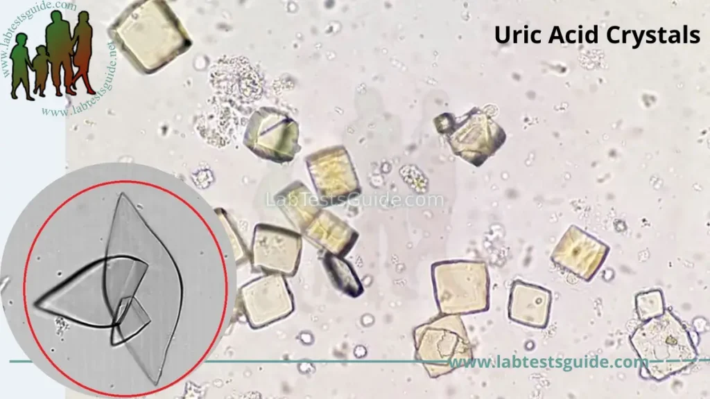
1. Uric acid Crystals:
Urate crystals can form when you have high levels of uric acid in your blood. Your body produces uric acid when it breaks down purines — substances that are found naturally in your body.
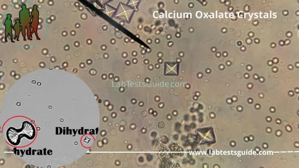
2. Calcium Oxalate Crystals:
Calcium oxalate crystals are found in individuals with acidic, neutral or alkaline urine. These crystals are colorless when viewed microscopically. There are two forms of the calcium oxalate crystal: the monohydrate and dihydrate form.
The monohydrate calcium oxalate crystal is described as the “picket fence” form. These dumbbells shaped crystals are common in ethylene glycol toxicity. The dihydrate form is octahedral or “envelope” shaped.
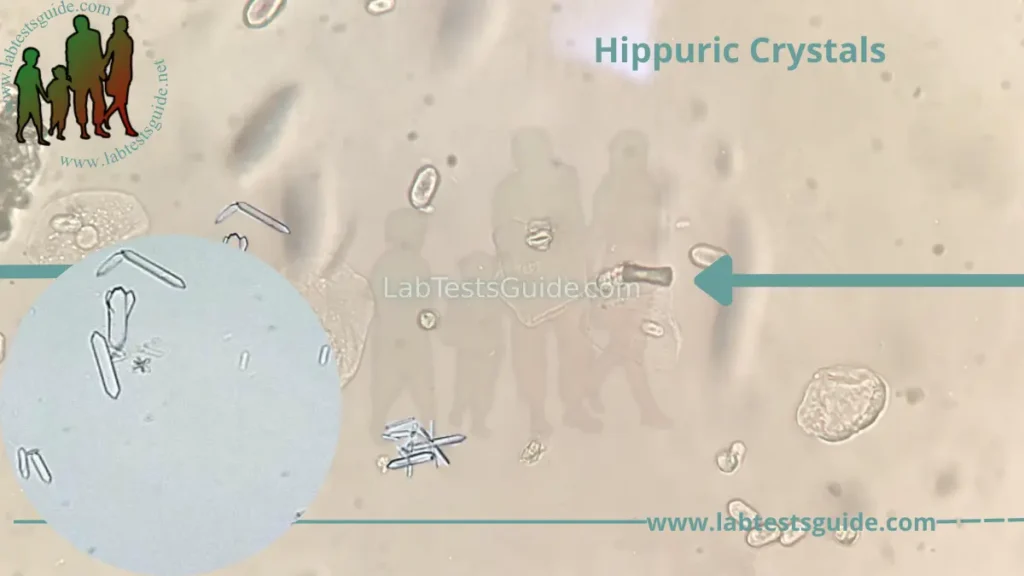
3. Hippuric Crystals :
Hippuric acid crystals are found in acid, neutral, or slightly alkaline urine. These colorless crystals are prisms, plates, or needle-like in shape. These crystals are often conglomerated into masses.
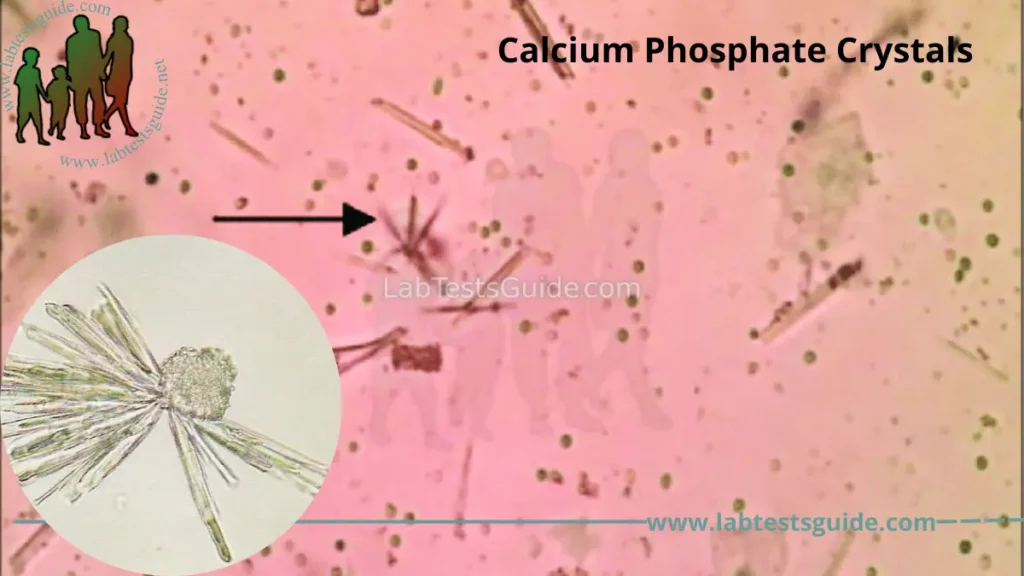
4. Calcium Phosphate Crystals :
These are colorless crystals having shape like blunt ended needles or prisms, rosettes. These crystals are found in neutral to alkaline pH.
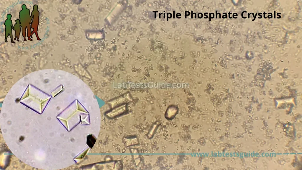
5. Triple Phosphate Crystals :
Triple phosphate crystals form in alkaline urine and are composed of magnesium, ammonium and phosphate. These are rectangular in shape or similar with the coffin lid. These are sometimes associated with a bacterial urinary tract infection caused by urea splitting bacteria.
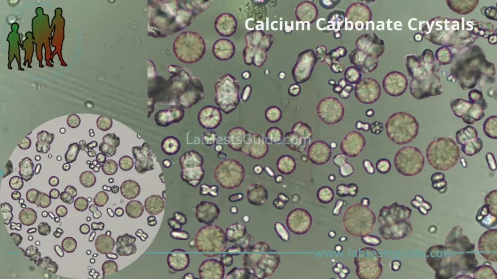
6. Calcium Carbonate Crystals :
Calcium carbonate crystals are yellow to colorless dumbells or spheres with radial striations, found in alkaline urine. They are usually large crystals and can be readily observed at low magnification.
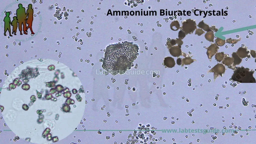
7. Ammonium Biurate Crystals:
Ammonium urate (or biurate) crystals generally appear as brown or yellow-brown spherical bodies with irregular protrusions resembling “throny-apples”. These are found in alkaline urine.
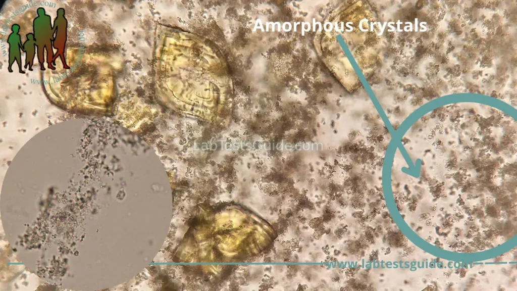
8. Amorphous Crystals:
Amorphous urates are found in acid urine. These crystals may appear pink on gross analysis and yellow microscopically. These crystals appear as granules in the urine sediment.
Amorphous phosphates are found in alkaline urine. These granules are colorless microscopically.
Abnormal Crystals
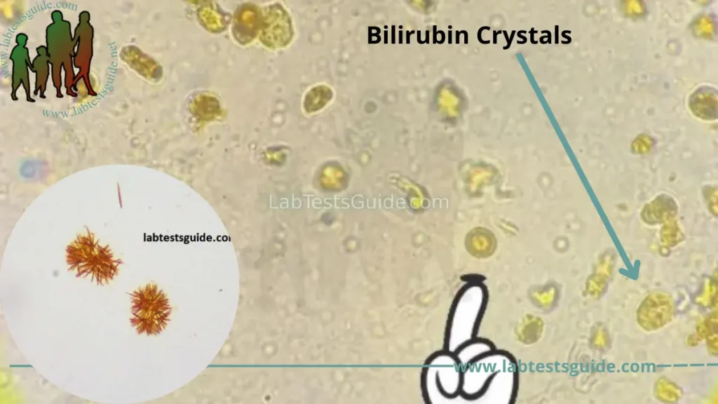
1. Bilirubin Crystals:
Bilirubin crystals are abnormal crystals in urine. They form from conjugated bilirubin and are needle-like to granular crystals that are yellow in color.They are frequently attached to the surface of cells. Bilirubin crystals are seen in several hepatic disorders.
2. Cholesterol Crystals :
These appear as colorless rectangular plates with a notch in one or more corners and are found in acidic urine. The appearance of cholesterol is associated with the Nephrotic Syndrome.

3. Cystine Crystals :
Cystine crystals are flat colorless plates and have a characteristic hexagonal shape with equal or unequal sides.
They occur in acidic urine that are associated with an inherited disorder. Presence of cystine crystals represents a proximal tubular defect in amino acid reabsorption.

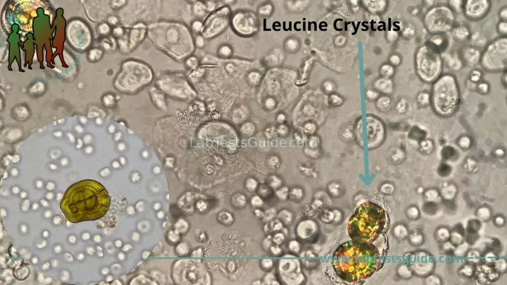
4. Leucine Crystals :
These are Yellowish-brown spheres with concentric circles with radial striations found in acidic/neutral urine. Leucine crystals may be seen in liver disorders in which amino acid metabolism is impaired.
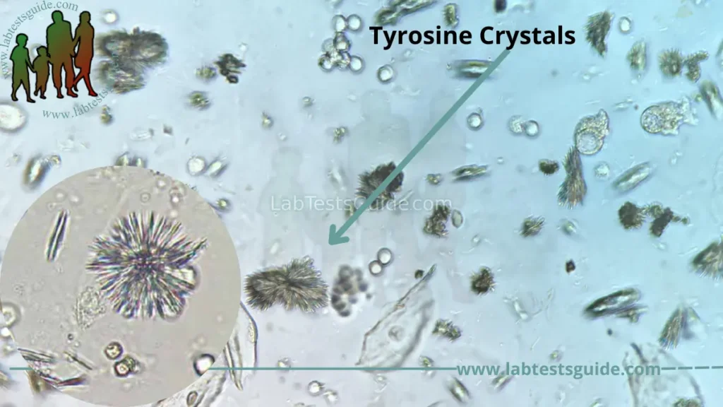
5. Tyrosine Crystals:
Tyrosine crystals appear as colorless/yellow fine needles in acidic/neutral urine. Tyrosine crystals may be seen in tyrosinemia and in certain liver disorders in which amino acid metabolism is impaired.
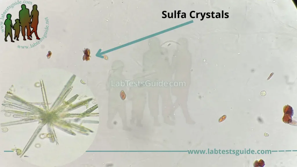
6. Sulfa Crystals:
These are flat needles, sheaves of small needles or as spheroids. Often brown in color. The presence of sulfanomide crystals usually indicates administration of the drug and not necessarily a pathological condition.
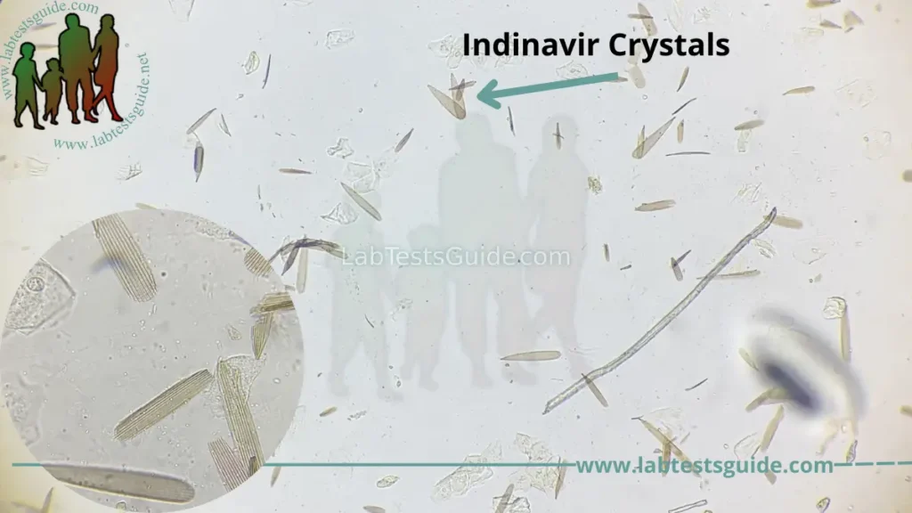
7. Indinavir Crystals :
Indinavir crystals can be observed readily in the urine sediment with the use of routine light microscopy. Approximately 20% of an oral indinavir dose is excreted in the urine during the ensuing 8 hours, half as the intact indinavir molecule and half as one or more of its seven known metabolites (three metabolites are closely related structurally to the parent compound).
Crystals in the acidic urine :
| Name of crystals | pH | Shape | Color |
|---|---|---|---|
| Sodium urate | acidic (<acidic) | amorphous | Amorphous, or large granules |
| Uric acid | acidic <5.5 | rhombic, four-sided flat plates | yellow-brown |
| Amorphous urates | acidic >5.5 | amorphous or sand like | Microscopically yellow-brown and occurs in clumps |
| Calcium oxalate | acidic or alkaline | enveloped shaped, dumbbell-shaped | The colorless octahedral envelope or two pyramids joined at the base. |
| Cystine | acidic | hexagonal | Colorless |
| Cholesterol | acidic | rectangular, notched plates | Colorless |
| Tyrosine | acid/neutral | needles shape form clumps or rosettes | Colorless to yellow, needles |
| Leucine | acidic /neutral | spheres with a concentric circle or radial striations | yellow-brown |
| Bilirubin | acid | clumped needles or granular | yellow color |
| Sulphonamide | acid/neutral | Rosette form, needle, | colorless to yellow-brown |
| Ampicillin | acid/neutral | in needles form | colorless |
| Radiographic dye | acid | like cholesterol | colorless |
Crystals in alkaline urine :
| Name of crystals | pH | Color and shape |
|---|---|---|
| Amorphous phosphates | Alkaline | Granular |
| Triple phosphates | Alkaline | Colorless and prism-shaped like a coffin lid |
| Calcium phosphate | Alkaline | Colorless, flat rectangular plates or thin prisms often in rosettes |
| Calcium Carbonate | Alkaline | Small, colorless with a dumbbell or spherical shape, may occur in clumps and resemble amorphous material |
| Ammonium biurate | Alkaline | Yellow-brown color, thorny apples |
Pathophysiology
- There may be well-defined crystals or amorphous material in the urine sediment.
- The presence of crystals in the urine is called Crystalluria.
- When urine left at room temperature or refrigerated then urine becomes cloudy because of precipitation of crystals or amorphous material.
- These crystals are important in case of kidney stones.
- The stone formation may be without crystals in the urine or crystalluria may be without stone formation.
- Some of the crystals indicate some metabolic disorders like cystinuria or sulfa drug.
- Crystals are seen mostly in the concentrated urine.
- Crystals are divided into :
- Normal or abnormal
- In alkaline or acidic urine.
- Crystals found due to medication.
- Crystals found in acidic urine has pH of <6.5 and in alkaline urine pH is >7.0.
Understand Your Test Results:
Understand your Urine Crystals Results
AI-powered Lab Test Results Meaning tool 🤖
20 FAQs:
What are crystals in urine?
Crystals in urine are solid structures that form from minerals present in the urine.
What are the different types of crystals found in urine?
The most common types of crystals found in urine include calcium oxalate, uric acid, struvite, and cystine.
What causes crystals to form in urine?
Crystals form in urine when there is an excess of minerals or a decrease in urine volume.
Are crystals in urine normal?
Small amounts of crystals in urine are normal and can be caused by factors like diet and dehydration. However, excessive amounts of crystals may be a sign of an underlying condition.
What are the symptoms of crystals in urine?
Most people with crystals in urine do not experience any symptoms. However, in some cases, it can cause pain during urination, frequent urination, and cloudy or foul-smelling urine.
Can crystals in urine cause kidney stones?
Yes, crystals in urine can sometimes clump together and form kidney stones.
How are crystals in urine detected?
Crystals in urine can be detected through a urine test or urinalysis.
How are crystals in urine treated?
Treatment depends on the underlying cause of the crystals. It may involve lifestyle changes, medication, or surgery.
Can diet affect the formation of crystals in urine?
Yes, certain foods and drinks can increase the likelihood of crystal formation, such as those high in calcium, oxalate, and purines.
What are the risk factors for developing crystals in urine?
Risk factors include dehydration, a diet high in certain minerals, obesity, and certain medical conditions like gout and kidney disease.
Can children develop crystals in urine?
Yes, children can develop crystals in urine.
Are there any complications associated with crystals in urine?
Complications can occur if the crystals clump together and form kidney stones, which can cause pain and other symptoms.
Can crystals in urine be prevented?
Prevention involves staying hydrated, maintaining a healthy diet, and treating underlying medical conditions.
How long do crystals in urine take to form?
The time it takes for crystals to form in urine depends on a variety of factors and can vary from person to person.
Can medications cause crystals in urine?
Yes, certain medications can cause crystals to form in urine.
Can crystals in urine go away on their own?
In some cases, small amounts of crystals can go away on their own without treatment.
Can crystals in urine be a sign of cancer?
In rare cases, crystals in urine can be a sign of bladder or kidney cancer.
Can exercise affect the formation of crystals in urine?
Exercise can help prevent the formation of crystals by increasing urine volume and promoting hydration.
Can stress affect the formation of crystals in urine?
Stress can indirectly affect the formation of crystals by causing dehydration and other changes in the body.
How often should I get my urine checked for crystals?
If you have a history of kidney stones or other urinary issues, your doctor may recommend regular urine tests to check for crystals. Otherwise, it may not be necessary to get your urine checked unless you experience symptoms.