

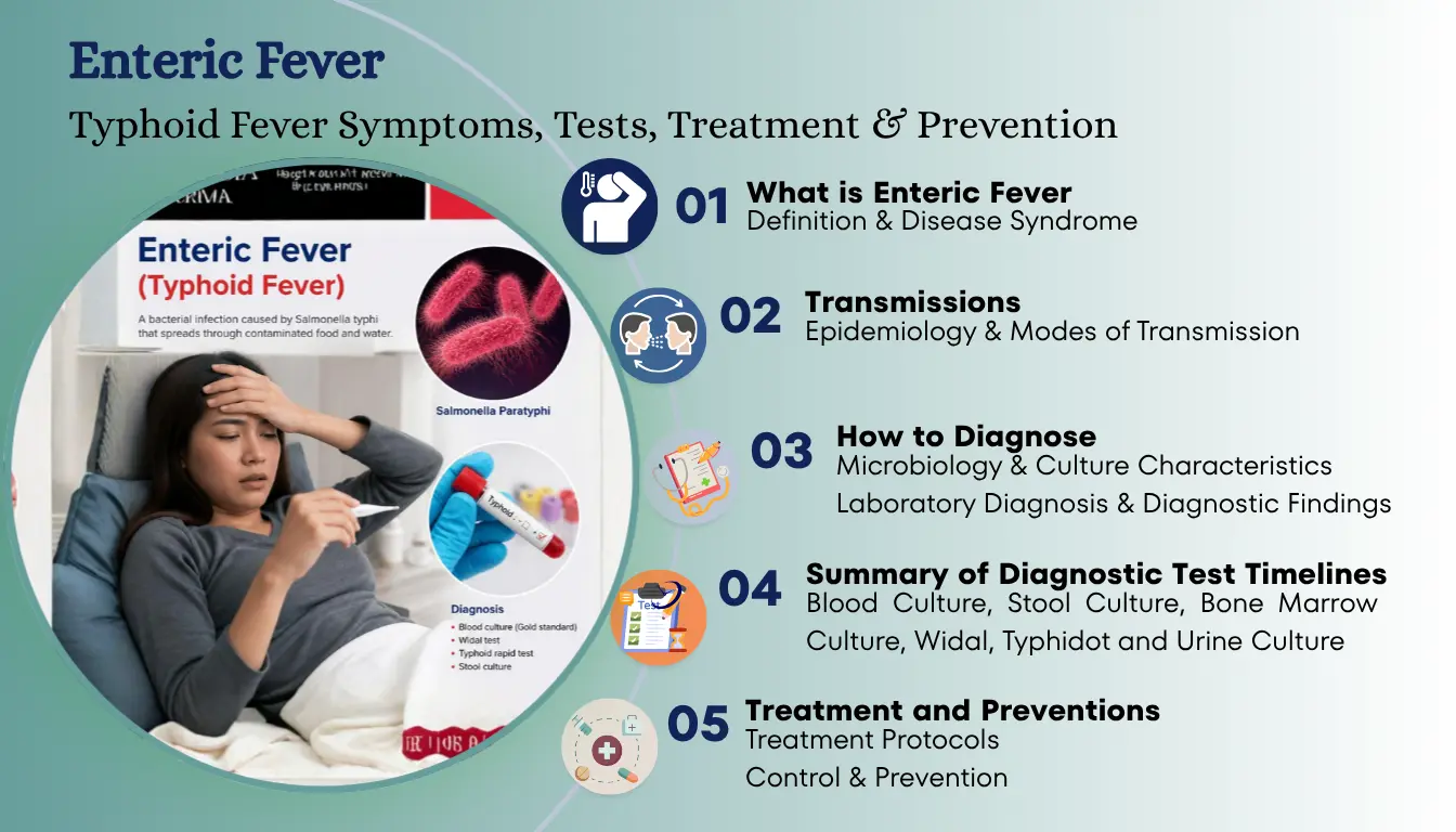
Enteric Fever: Typhoid Fever Symptoms, Tests, Treatment & Prevention
Definition & Disease Syndrome
Typhoid fever (also known as Enteric Fever) is caused by infection with Salmonella typhi and Salmonella paratyphi.
Salmonella typhi is found only in humans; no animal reservoir has been identified.
Typhoid/Enteric fever is a clinical syndrome characterized by:
- Fever
- Headache
- Cough
- Splenomegaly
- Leucopenia
Important Facts & Infections Division
Typhoid fever is an acute illness characterized by fever, headache, and abdominal discomfort. Salmonella infections are categorized into:
1. Typhoidal Infection:
- Caused by S. typhi.
- S. paratyphi generally causes a less severe illness (types A, B, or C).
2. Non-Typhoidal Infection:
- Most common in the USA, caused by S. Typhimurium and S. Enteritidis.
- Salmonella is uncommon in the USA; 70% are acquired abroad.
Humans are the only reservoirs of S. typhi and S. paratyphi. Transmission occurs person-to-person or via contaminated food or water.
Epidemiology & Modes of Transmission
Transmission Factors:
- Salmonella habitat is the animal gut. Predominantly animal pathogens can also cause disease in humans.
- Foodstuffs of animal origin are important vehicles for transmission.
- Found in humans as well as in chickens and eggs. Spreads by eating chickens and uncooked eggs.
- Spreads through close contact with an acutely infected person or chronic carrier.
- Most cases occur through contaminated foods or water.
Global Epidemiology & Risk Demographics:
- Global issue: 13 to 17 million cases and ~60,000 deaths yearly.
- Children under 1 year are more prone to developing infection, and diseases are more severe.
- There is increasing antibiotic resistance to this disease.
- Salmonella typhi and S. paratyphi differ from other species because humans are the only natural hosts.
Microbiology & Culture Characteristics
Microbiological Profile: Gram-negative bacilli (rods), seen as single pairs and chains. Aerobic, motile (via flagella), non-lactose fermenters, and produce H2S gas. Capsules consist of polysaccharides. Colonies are gray and grow on ordinary media (blood agar).
Classification & Differentiation:
Salmonella is serologically divided into: (1) Salmonella typhi, (2) Salmonella cholerae-suis, (3) Salmonella enteritidis.
Other Salmonella types live in the GIT of animals and infect humans when food/water is contaminated with animal feces. S. typhi grows on ordinary media; unlike most salmonellae that produce acid and gas from carbohydrates, S. typhi does not produce gas.
Culture Media Observations:
- MacConkey Agar / Desoxycholate Citrate Agar: Pale, non-lactose fermenting colonies (from feces).
- Xylose Lysine Desoxycholate (XLD) Agar: Red colonies with a central black area.
- Wilson & Blair’s Bismuth Sulfite Agar: Black metallic colonies due to H2S production.
- Enrichment Media: Selenite F and tetrathionate broth for enrichment, then subculture to MacConkey agar.
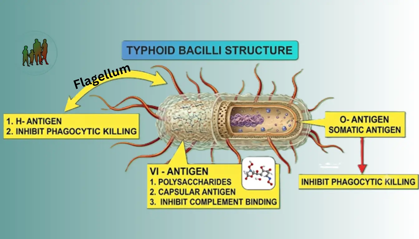
Salmonella Antigens
1. O – Antigen (Somatic):
- Present in bacterial body.
- Endotoxin; provides antigenic variation.
- Thermostable & resists alcohol extraction.
2. H – Antigen (Flagellar):
- Present in flagella; provides motility.
- Undergoes variation via gene rearrangement.
3. Vi – Antigen (Capsular/Envelope):
- Superficial capsular polysaccharide (“Vi” = Virulence).
- Surrounds O antigen, protecting from antibody/complement attack.
Respective Antibodies: Anti-O, Anti-H, and Anti-Vi antibodies.
Pathology & Pathogenesis
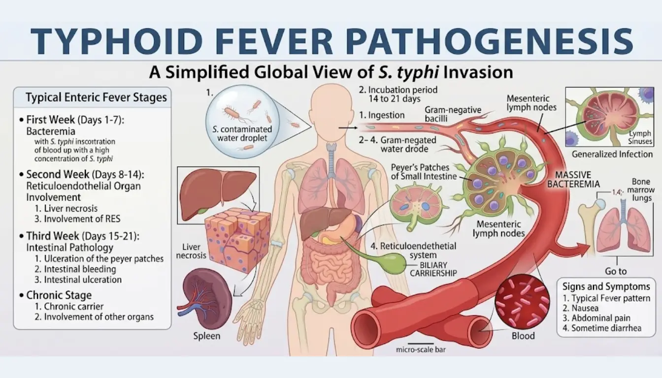
Three Primary Disease Manifestations: (1) Enteric fever, (2) Gastroenteritis, (3) Septicemia.
Incubation Periods: Typhoid fever: 5 to 14 days (range: 1–3 weeks / 3–60 days). Salmonella gastroenteritis: 6 to 72 hours.
Pathogenic Sequence:
- Organism crosses intestinal epithelium and invades regional lymph nodes.
- Multiplies inside macrophages of Peyer’s patches, mesenteric lymph nodes, and spleen.
- Peyer’s patches become inflamed and may ulcerate.
- Bacteremia occurs; infection spreads to lungs, gallbladder, kidneys, and CNS, settling in organs.
- Phagocytosed by monocytes (mononuclear phagocytes), surviving intracellularly as facultative intracellular parasites.
Note: S. typhi and S. paratyphi A, B, C cause enteric fever. Other species cause gastroenteritis/food poisoning or septicemia. Rarely causes osteomyelitis, septic arthritis, and purulent lesions.
Stages of Salmonella / Enteric Fever Progression:
Insidious onset, bacteremia, rising stepwise fever, headache, abdominal pain, malaise.
Splenomegaly, widespread reticuloendothelial involvement, necrotic foci in liver, peak fever.
Peyer’s patches ulcerate; risks of intestinal ulcers, severe bleeding, and perforation.
Colonizes gallbladder, potentially leading to chronic carrier state or organ lesions.
1. Prodromal & Systemic Stage
Clinical Manifestations:
- Four Disease Types: Typhoid fever, Sepsis, Gastroenteritis (diarrhea), Carrier state.
- Demographics: Infection is more common in children under 10 years of age. Relapse occurs in 5% to 10% of untreated cases.
- Initial Symptoms: Malaise, headache, cough, sore throat, abdominal pain, constipation. Diarrhea is a late symptom (usually 3rd week).
- Fever Pattern: Constant and ascends in a stepwise pattern. Rises in 1st week, peaks in 2nd, decreases in 3rd.
- 7 to 10 Days: Peak fever, patients are sicker; early diarrhea, abdominal distension, and cramps.
- Recovery: Patients improve after 7–10 days. Untreated cases recover in ~4 weeks. Symptoms disappear with antibiotics within 3–4 days. Relapse may occur after 2 weeks.
Key Signs & Symptoms: Early abdominal tenderness, hepatosplenomegaly, and scanty maculopapular rash (rose spots).
2. Gastroenteritis Stage
Clinical Manifestations:
- Incubation Period: 8 to 24 hours.
- Symptoms: Nausea, vomiting, abdominal pain, and fever.
- Diarrhea Characteristics: Stools are loose, moderate volume, and do NOT contain blood.
- Duration: Diarrhea is self-limited, lasting 3 to 7 days. Self-limiting disease with recovery usually within a week.
- Differential Diagnosis: Signs/symptoms may mimic appendicitis or inflammatory bowel disease (IBD).
Complications & Chronic Carrier State
Systemic & Late Complications:
Usually occur after the 3rd week of disease (untreated or treated):
- Intestinal Hemorrhage: Occurs in 20% of cases (usually 2nd/3rd week); less common in treated cases.
- Intestinal Perforation: Occurs in 3% of untreated patients.
- Relapse: Occurs in 1 in 5 patients (10% untreated; 5-10% general).
- Organ Involvement: Meningitis, Typhoid abscesses (rare), Lobar pneumonia, Myocarditis, Osteomyelitis, Periostitis (rare), Bacteremia/Sepsis (infecting lungs, brain, bones).
- Secondary Suppurative Lesions: Lungs (pneumonia), parotid gland (parotitis), furunculosis.
- Mortality/Outcome: 4th week improvement seen, but 30% mortality in developing countries without treatment (1-2% in USA).
- Lab Changes in Complications: Increased LDH, Alkaline Phosphatase, SGOT, CPK, and abnormal liver function tests (LFTs).
The Carrier State Definition & Features:
- ~30% of patients become carriers due to persistent gallbladder infection.
- Convalescent Carriers: 5% to 10% excrete bacteria for several months post-recovery.
- Chronic Carriers: 1% to 4% carry organisms for >1 year.
- Carriers have no active disease and are symptom-free.
- Bacteria from gallbladder discharge constantly into intestine → feces. Associated with cholecystitis and gallstones.
- Difficult to eradicate. Requires high-dose prolonged antibiotics or surgical cholecystectomy.
Laboratory Diagnosis & Diagnostic Findings
CBC & Hematology:
- WBC: Low TLC / Leucopenia.
- Weeks 1–2: 4,000–6,000 /cmm.
- Weeks 3–4: 3,000–5,000 /cmm.
- >10,000 /cmm suggests perforation or suppuration.
- ESR: Usually decreased.
- Anemia: Normocytic anemia; becomes hypochromic microcytic if bleeding occurs.
Culture Protocols (Gold Standard):
- Blood Culture: >15 mL blood. 80–90% positive in 1st 10 days; <30–50% by 3rd week. 100% specific. Count required: 105–109 organisms (lower in infants, elderly, H2 blockers/antacids users). Buffy coat reduces isolation time.
- Bone Marrow Culture: Most sensitive (80–95% / up to 90%), regardless of stage or prior antibiotics.
- Stool Culture: Positive after 7–10 days; peak sensitivity (<50%) in 4th–5th week. Positive >4 months = carrier (3%). Shows leucocytes, rarely RBCs.
- Urine Culture: Positive 2nd–3rd week in 25% cases (even if blood culture negative). Done on MacConkey agar.
Serology & Rapid Tests:
- Biochemical Profile: Oxidase (-), Indole (-), Lactose (-), Urease (-), VP (-), Citrate (+/-), TSI (+ H2S, except S. typhi gas -).
- Serogroups: 5 groups (A, B, C, D, E) via O-antigen; 1200 serotypes via H-antigen. Anti-D appears in 7–10 days.
- Typhidot Test: Claims positivity within 2 to 3 days.
- Phage Typing: Used for epidemiologic identification of strains.
- Vi Antigen: Screens carriers (positive in 70% chronic carriers).
Characteristic Biochemical Features on Culture:
| Lab Test | Result | Lab Test | Result |
|---|---|---|---|
| Oxidase Test | Negative | Voges-Proskauer | Negative |
| Indole Test | Negative | Simmon’s Citrate | Positive / Negative |
| Lactose Fermentation | Negative (Non-lactose) | TSI (H2S Production) | Positive (S. typhi No Gas) |
| Urease Test | Negative | Motility | Positive (Flagella) |
Widal Test Evaluation & Drawbacks
Widal Test Interpretation (Developed 1896):
Positive after 7 to 10 days. Useful only if culture facilities are unavailable.
- O-Antibodies (Somatic): Indicates acute infection; rises early, peaks at 3–5 weeks.
- 1:40 = Negative
- 1:80 = Suspicious / Borderline (Unvaccinated). Repeat in 5–7 days.
- 1:160 = Strongly suggestive of infection.
- H-Antibodies (Flagellar): Appears later, lasts longer; indicates past infection/vaccination.
- 1:40 = Suspicious (Unvaccinated).
- 1:160 = Strongly suggestive.
- Titers are significantly higher in vaccinated individuals.
Drawbacks & Limitations of Widal Test:
- Absent in early illness (<7 days).
- Antibody response is variable and does NOT correlate with disease severity.
- Early antibiotic therapy prevents titer rise or keeps it low/negative.
- Inadequate O-antibody response in some patients.
- False positives occur due to vaccination, past infection, or autoimmune diseases.
- >10% of cases in endemic areas are seronegative.
- Blood, bone marrow, bile, and stool cultures are superior.
Summary of Diagnostic Test Timelines
| Diagnostic Modality | Optimal Timeframe | Sensitivity / Specificity | Key Diagnostic Clinical Notes |
|---|---|---|---|
| Blood Culture | First 7–10 days (Week 1) | 80–90% sensitive, 100% specific | Requires >15 mL blood. Drops to <30-50% by Week 3. Positive in relapse. |
| Bone Marrow Culture | All stages (Weeks 1–4) | 80–95% sensitive | Most sensitive test; unaffected by prior antibiotic therapy. |
| Stool Culture | Weeks 3–5 (>10th day) | <50% positive | Positive >4 months indicates chronic carrier state (3%). |
| Urine Culture | Weeks 2–3 | 25% sensitive | Useful when blood culture is negative. Performed on MacConkey. |
| Widal Serology | After 7–10 days (Peak Wk 3-5) | Unreliable / Low Specificity | 4-fold rise in O titer. Affected by prior vaccines and antibiotics. |
| Typhidot | Early (Days 2–3) | Rapid Qualitative | Detects specific IgM/IgG antibodies early in disease. |
Treatment Protocols
Acute Enteric Fever Treatment:
- Drug of Choice: Ciprofloxacin
- Chloramphenicol: Highly effective, but carries serious potential side effects.
- Co-trimoxazole: Effective alternative with fewer serious side effects than Chloramphenicol.
Chronic Carrier Management:
- Asymptomatic carriers shed S. typhi in stool for >1 year (1–3% of cases). Gallbladder is primary site.
- Ampicillin: Drug of choice for carriers; must be administered in high doses for prolonged duration.
- Ciprofloxacin: Demonstrates good clinical eradication results.
- Chloramphenicol is NOT suitable for treating carriers.
- Surgical Intervention: Cholecystectomy (gallbladder removal) required in refractory permanent carriers.
Control & Prevention
Hygienic Measures:
- Clean municipal water supply and adequate sewage disposal.
- Proper food handling, processing, and hygiene.
- Handwashing: Thorough handwashing after defecation (wash hands with soap at least 3 times).
- Carriers must receive treatment and strict handwashing counseling.
- Vaccination recommended for families with history of enteric fever.
Vaccine Comparison:
| Vaccine Type | Dose & Admin | Remarks & Adverse Effects |
|---|---|---|
| Vi Capsular Polysaccharide (ViCPS – Typhim Vi) | 25 µg in 0.5 mL I/M single dose. Booster: Every 2 yrs. | Not for <2 yrs. Side effects: Fever (1%), Headache (1.5–3%), Local Erythema (7%). |
| Heat-Killed Whole Organism (S. typhi) | 0.5 mL I/M (2 doses, 4 wks apart). Booster: 0.1 mL q3yrs. (Kids 0.5–10 yrs: 0.25 mL) | High side effects: Fever (17–29%), Severe Headache (10%), Injection Site Pain (35–60%). |
| Ty21a (Vivotif Berna) | Live oral attenuated. 1 cap 1hr before meal q2d (4 days total). Booster: Every 5 yrs. | Few side effects. Contraindicated in immunocompromised & children <6 years. |
Layman’s Guide & Patient Communication
Understanding Widal Test Results for Patients:
- The Widal test becomes positive only after 7 to 10 days of fever onset.
- O-Antigen: Indicates an acute (active) infection and rises early.
- H-Antigen: Appears later than O-antigen; indicates past infection or previous vaccination.
- Titer Readings:
1:40= Negative.1:80= Borderline/Suspicious. Re-test in 5 to 7 days. A rising titer confirms typhoid fever. If levels stay identical, typhoid is unlikely.1:160= Strongly indicates active typhoid infection.
- Early Testing Advice: Widal test is negative during days 1 to 7. For fever in the first few days, order a Blood Culture instead.
Frequently Asked Questions (Clinical FAQs)
Blood culture is up to 90% sensitive and 100% specific during the first 7–10 days of infection because bacteremia is highest during this prodromal phase. In contrast, Widal antibodies (O and H agglutinins) take 7 to 10 days to reach detectable levels in serum, leading to false-negative Widal results in the first week.
About 1% to 4% of patients become chronic carriers when Salmonella typhi persistently colonizes the gallbladder (often associated with gallstones or cholecystitis). Organisms are continuously shed into the stool for over a year. Treatment requires prolonged high-dose antibiotics (Ampicillin or Ciprofloxacin). In resistant cases, surgical removal of the gallbladder (cholecystectomy) is necessary.
Both are forms of enteric fever transmitted via contaminated food/water. Typhoid fever is caused by Salmonella typhi, whereas Paratyphoid fever is caused by Salmonella paratyphi (types A, B, or C). Paratyphoid fever generally presents as a milder clinical illness than typhoid fever.
Major complications usually occur during the 3rd week of illness and include Gastrointestinal Hemorrhage (occurs in ~20% of untreated cases) and Intestinal Perforation (occurs in ~3% of untreated cases) secondary to necrosis of ulcerated Peyer’s patches. Other severe risks include myocarditis, lobar pneumonia, and severe sepsis.
The Vi Capsular Polysaccharide (ViCPS) injectable vaccine is widely used for individuals aged 2 years and older (requires a booster every 2 years). The Ty21a oral live attenuated vaccine is suitable for adults and children over 6 years old (given as 4 capsules over 7 days, booster every 5 years), but it must NOT be given to immunocompromised individuals.
Salmonella typhi lacks the enzymes necessary to ferment lactose. As a result, when grown on MacConkey agar, it does not produce acid to change the neutral red indicator, forming pale, colorless, non-lactose fermenting colonies.
References and Sources
- Typhoid Fever: Causes, Symptoms & Treatment – Cleveland Clinic – (accessed on July 20, 2026)
- Typhoid Fever: Background, Etiology, Pathophysiology – Medicine – Medscape – (accessed on July 20, 2026)
- Typhoid fever – A Comprehensive Guide – Wikipedia – (accessed on July 20, 2026)
- Enteric Fever Diagnosis: Current Challenges and Future Directions – PMC – National Library of Medicine – NIH – (accessed on July 20, 2026)
- Typhoid Fact Sheed by World Health Organisation (WHO) – WHO – (accessed on July 20, 2026)
- Typhoid fever – Diagnosis & treatment – Mayo Clinic – Mayo Clinic Lab – (accessed on July 20, 2026)
- Enteric Fever:- Part (1-4) – Typhoid Fever (Salmonella typhi), Diagnosis and Treatment – LabPedia.net – (accessed on July 20, 2026)
