Anti TTG Antibodies Test Purpose, Procedure, Principle, Result Interpretation, Report Formate and Clinical Signification
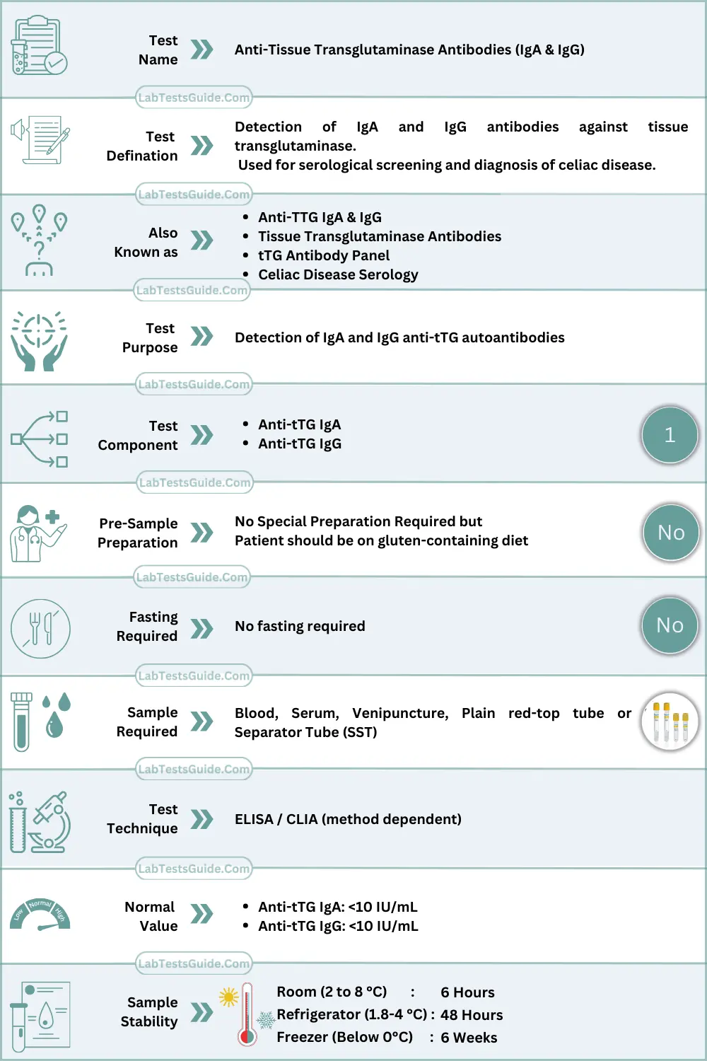
The Anti-tTG antibody assay is an immunological serological test used to detect autoantibodies directed against tissue transglutaminase (tTG), an intracellular enzyme involved in post-translational protein modification. The test is primarily utilized for autoimmune enteropathy assessment, especially gluten-related immune disorders.
The assay may detect IgA and/or IgG class antibodies, depending on the test configuration.

🧠 AI-Powered Test Result Analysis:
Understand your Anti-Tissue Transglutaminase (Anti-tTG) Antibodies Test Results
AI-powered Lab Test Results Meaning tool 🤖
Anti TTG Test Types:
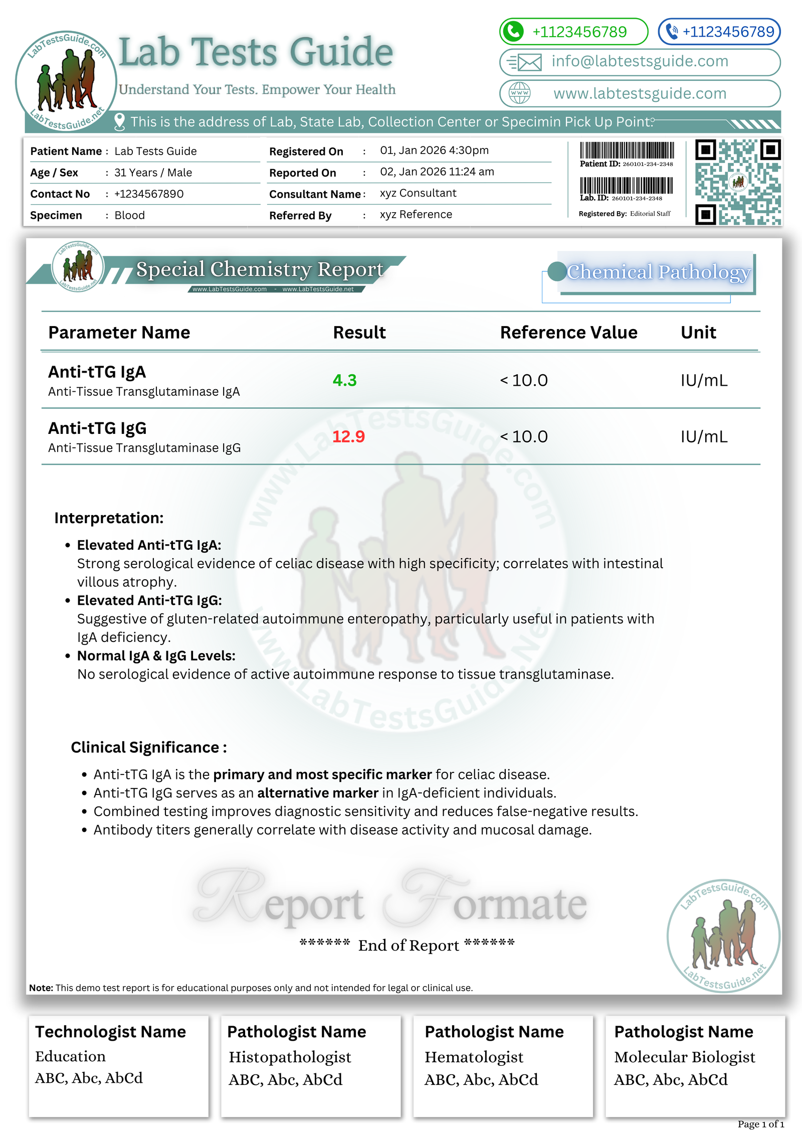
- Anti-TTG IgA Test:
- This test measures IgA (immunoglobulin A) antibodies against tissue transglutaminase.
- IgA antibodies are commonly used because they are found in the mucous membranes of the gastrointestinal tract, which is where celiac disease primarily affects.
- It is the primary form of the Anti-TTG antibody test and is considered highly specific for celiac disease diagnosis.
- Results are typically reported in units such as U/mL (units per milliliter) or IU/mL (international units per milliliter).
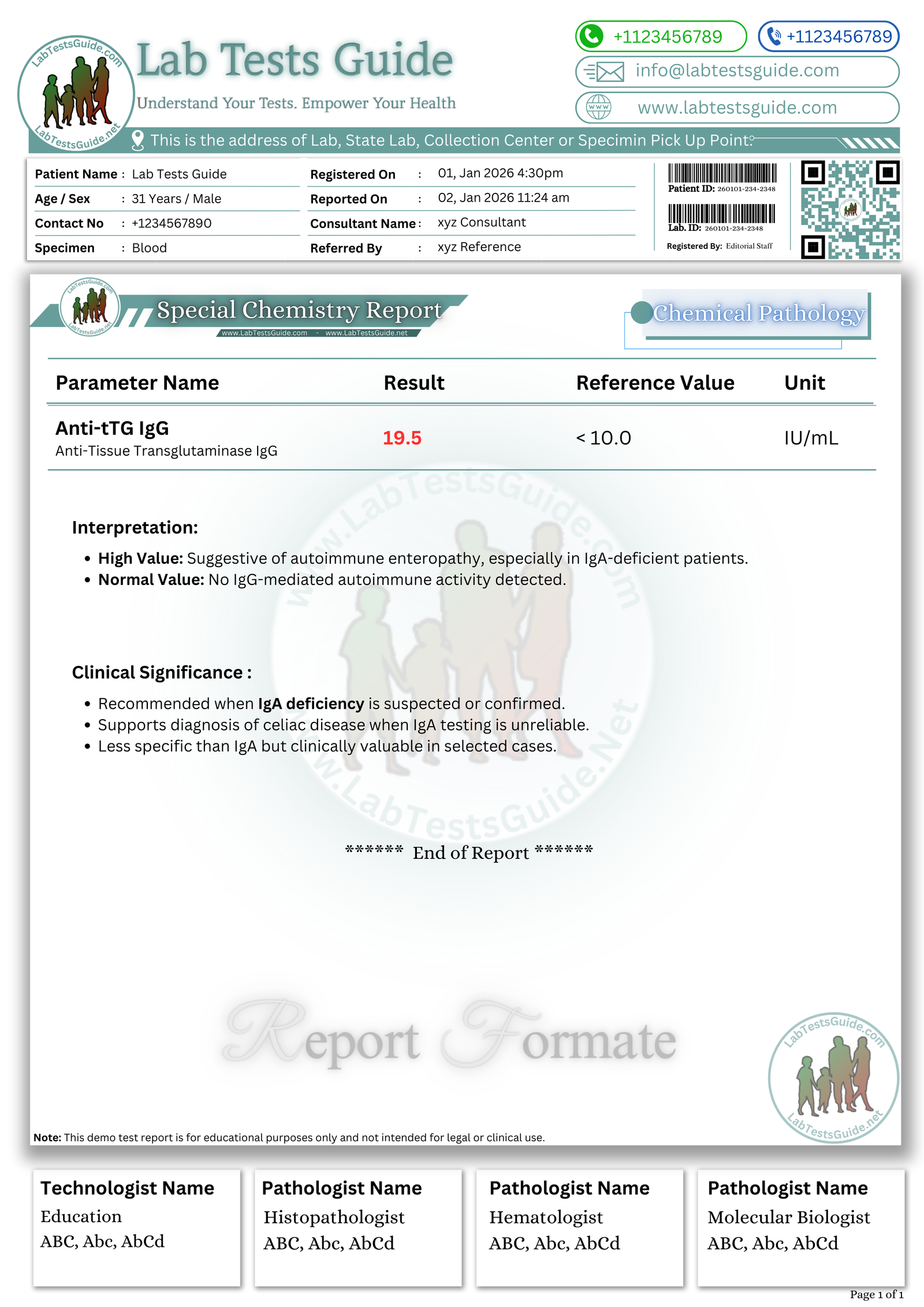
- Anti-TTG IgG Test:
- In some cases, individuals with celiac disease may have IgA deficiency, which can lead to false-negative results on the Anti-TTG IgA test.
- In such cases, the Anti-TTG IgG test is used to detect IgG antibodies against tissue transglutaminase.
- This test is often used as an alternative when IgA deficiency is suspected or confirmed.
- Results are also reported in units such as U/mL or IU/mL.

Principle of the Test
The Anti-tTG assay is based on an antigen–antibody immunological reaction.
Method Type:
- Enzyme-Linked Immunosorbent Assay (ELISA) – most common
- Chemiluminescent Immunoassay (CLIA)
- Fluorescence Enzyme Immunoassay (FEIA)
- Radioimmunoassay (RIA) – obsolete/rare
ELISA Principle
- Microtiter wells are coated with recombinant human tissue transglutaminase antigen
- Patient serum antibodies (IgA or IgG) bind to immobilized tTG
- Enzyme-labeled anti-human Ig conjugate binds to antigen-antibody complexes
- Substrate (e.g., TMB) reacts with enzyme producing a color change
- Color intensity is directly proportional to antibody concentration
- Measured photometrically at 450 nm
Clinical Significance (Technical)
Elevated Anti-tTG Levels
- Indicates autoimmune-mediated mucosal injury
- Reflects loss of immune tolerance to tissue transglutaminase
- Strong association with:
- Gluten-mediated autoimmune enteropathy
- Malabsorption syndromes
- High titers correlate with intestinal villous atrophy severity
- IgA Anti-tTG shows highest diagnostic accuracy
- IgG Anti-tTG is significant in IgA-deficient states
Low or Undetectable Levels
- Suggest absence of autoimmune response
- May occur in:
- Early disease
- Immunoglobulin deficiencies (e.g., selective IgA deficiency)
- Requires correlation with total serum IgA
Specimen Requirements
| Parameter | Requirement |
|---|---|
| Specimen Type | Serum (preferred) |
| Acceptable Specimen | Plasma (EDTA or heparin – method dependent) |
| Tube Color | Red, Yellow (SST) |
| Minimum Volume | 0.5 mL serum |
| Additives | None (serum preferred) |
| Stability | 48 hours at 2–8 °C |
| Long-term Storage | −20 °C (≤3 months) |
| Transport | Cold chain recommended |
| Rejection Criteria | Hemolysis, lipemia, microbial contamination, delayed separation, incorrect tube |
Patient Preparation (Technical)
- No fasting required
- Sample should be collected before immunosuppressive therapy
- Ongoing gluten exposure may affect antibody levels
- Avoid repeated freeze-thaw cycles
- Recent blood transfusion may interfere
Reagents & Materials Required
Reagents
- Recombinant human tTG-coated microplates
- Enzyme-labeled anti-human IgA or IgG conjugate
- TMB substrate
- Stop solution (0.5–1N sulfuric acid)
- Wash buffer concentrate
Calibrators
- Multi-level calibrators traceable to manufacturer standard
Controls
- Negative control
- Low positive control
- High positive control
Equipment
- Micropipettes (10–1000 µL)
- ELISA washer
- Microplate reader
- Incubator (37 °C)
- Timer
Wavelength
- Primary: 450 nm
- Reference: 620–630 nm (optional)
Reagent Storage
- 2–8 °C
- Do not freeze coated plates
Test Procedure (Step-by-Step)
A. Manual ELISA Method
- Bring reagents and samples to room temperature
- Add 100 µL calibrators, controls, and samples to wells
- Incubate at 37 °C for 30 minutes
- Wash wells 3–5 times
- Add 100 µL enzyme conjugate
- Incubate at 37 °C for 30 minutes
- Wash thoroughly
- Add 100 µL TMB substrate
- Incubate 10–15 minutes in dark
- Add 100 µL stop solution
- Read absorbance at 450 nm
B. Automated Method
- Load samples, reagents, and calibrators
- Program assay protocol
- Perform calibration as per analyzer instructions
- Analyzer performs incubation, washing, detection
- On-board reagent stability: 7–14 days (method dependent)
Calculation Formula
Manual ELISA Calculation
Anti-tTG (IU/mL)=Calibrator AbsorbanceSample Absorbance×Calibrator Value
Units: IU/mL
Example:
Sample OD = 1.20
Calibrator OD = 0.80
Calibrator = 10 IU/mL
Result = (1.20 / 0.80) × 10 = 15 IU/mL
Reference Ranges
| Population | Reference Range (IU/mL) |
|---|---|
| Adult Male | <10 |
| Adult Female | <10 |
| Children | <10 |
| Pregnancy | <10 |
| Method-specific | As per manufacturer |
Note: Reference ranges must be validated by each laboratory.
Interpretation (Technical / Professional)
High Levels
- Autoimmune antibody production
- Active mucosal immune injury
- Correlates with:
- Anti-endomysial antibodies
- Elevated inflammatory markers
- Malabsorption biochemical profile
Low Levels
- Absence of autoimmune activity
- Possible IgA deficiency
- Early disease or remission state
Interfering Factors / Sources of Error
- Hemolysis → false elevation
- Lipemia → optical interference
- Icterus → absorbance distortion
- High bilirubin
- Immunosuppressive drugs ↓ levels
- Improper washing
- Incorrect anticoagulant
- Temperature fluctuations
- Instrument drift
- Microbial contamination
Quality Control Requirements
- QC Levels: Normal, Low Positive, High Positive
- Frequency: Daily or per batch
- Rules: Westgard 1-2s, 2-2s, R-4s
- Charts: Levey–Jennings mandatory
- Corrective Action: Recalibrate, repeat run, check reagents
Calibration Requirements
- Multi-level calibration
- With new reagent lot
- After analyzer maintenance
- Linearity typically up to 200 IU/mL
- Recalibrate if QC fails
Instrument Maintenance Notes
- Daily probe washing
- Weekly optical cleaning
- Monthly deep wash cycles
- Check temperature calibration
- Prevent carryover contamination
🧠 AI-Powered Test Result Analysis:
Understand your Anti-Tissue Transglutaminase (Anti-tTG) Antibodies Test Results
AI-powered Lab Test Results Meaning tool 🤖
📥 Download Anti TTG Antibodies Test Report Format
Get the demo report format for Electrolytes Panel in your preferred format. These templates are fully editable and professional.
How to Download ?
| File Description | Format |
|---|---|
Anti-tTG Antibodies Test Report Format (Image) | .PNG ⬇️ |
Anti-tTG Antibodies Test Report Format (MS Word) | .DOCX ⬇️ |
Anti-tTG Antibodies Test Format (MS Excel) | .XLSX ⬇️ |
Anti-tTG Antibodies Test Format (PDF) | .PDF ⬇️ |
Anti-tTG IgA Test Report Format (Image) | .PNG ⬇️ |
Anti-tTG IgA Test Report Format (MS Word) | .DOCX ⬇️ |
Anti-tTG IgA Test Format (MS Excel) | .XLSX ⬇️ |
Anti-tTG IgA Test Format (PDF) | .PDF ⬇️ |
Anti-tTG IgG Test Report Format (Image) | .PNG ⬇️ |
Anti-tTG IgG Test Report Format (MS Word) | .DOCX ⬇️ |
Anti-tTG IgG Test Format (MS Excel) | .XLSX ⬇️ |
Anti-tTG IgG Test Format (PDF) | .PDF ⬇️ |
Anti-tTG IgG & IgA Test Report Format (Image) | .PNG ⬇️ |
Anti-tTG IgG & IgA Test Report Format (MS Word) | .DOCX ⬇️ |
Anti-tTG IgG & IgA Test Format (MS Excel) | .XLSX ⬇️ |
Anti-tTG IgG & IgA Test Format (PDF) | .PDF ⬇️ |
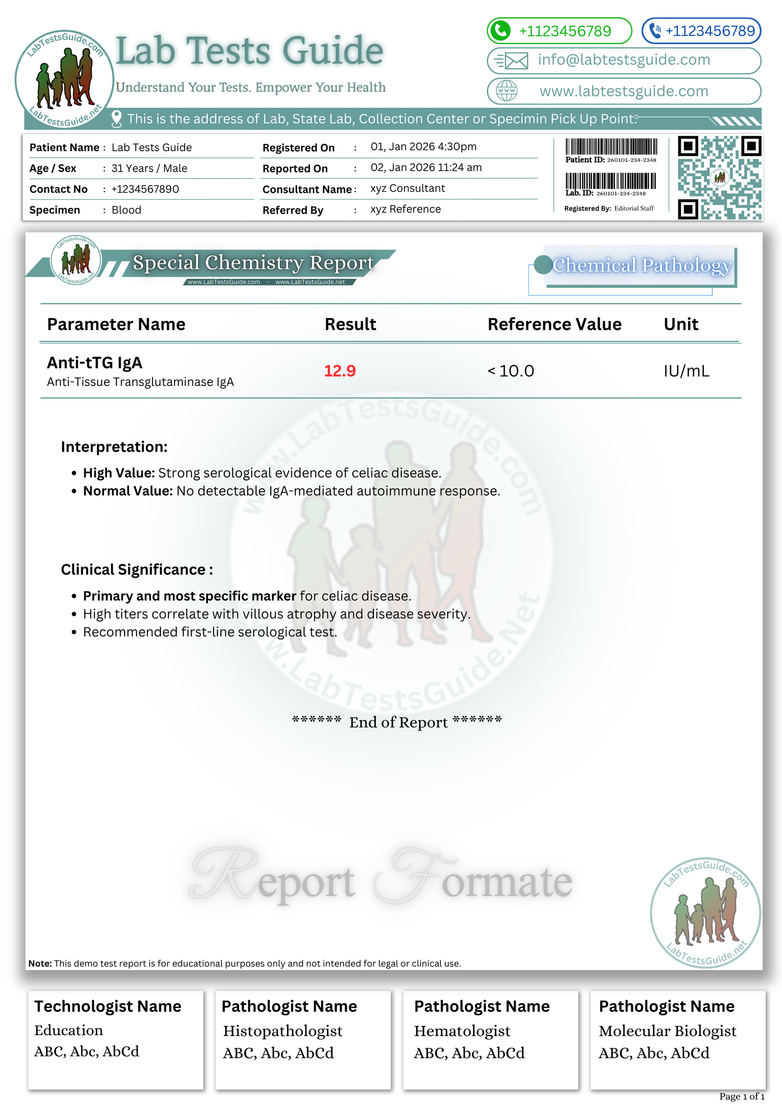
Reporting Format
Test: Anti-tTG Antibodies (IgA)
Result: 18 IU/mL
Reference Range: <10 IU/mL
Flag: H
Method: ELISA
Technical Note: Results should be interpreted with total IgA levels.
Critical (Panic) Values
- 100 IU/mL (method-dependent)
Follow institutional policy for notification.
Troubleshooting Guide
| Problem | Cause | Solution |
|---|---|---|
| Low absorbance | Expired reagent | Replace reagent |
| High blank | Contamination | Prepare fresh substrate |
| Drift | Temperature issue | Re-equilibrate analyzer |
| QC failure | Calibration error | Recalibrate |
| Sample clot | Improper centrifugation | Re-collect |
Safety Precautions
- Use PPE (gloves, lab coat)
- Treat all samples as biohazard
- Dispose reagents per MSDS
- Avoid aerosol formation
- Decontaminate spills immediately
Test Limitations
- False negatives in IgA deficiency
- Cross-reactivity with other autoimmune antibodies
- Not suitable for monitoring acute changes
- Method-dependent sensitivity variations
Notes for Lab Students
- Always check total IgA status
- Strict washing steps are critical
- QC validation is mandatory
- Never report without calibration verification
Viva / MCQs
- Anti-tTG detects antibodies against which enzyme?
- Primary wavelength used in ELISA?
- Most common specimen type?
- Main cause of false low results?
- Which QC rule detects random error?
Answers:
- Tissue transglutaminase
- 450 nm
- Serum
- IgA deficiency
- R-4s rule
FAQs:
What is the Anti-TTG antibodies test?
The Anti-TTG antibodies test is a blood test used to detect the presence and quantity of antibodies against tissue transglutaminase. It is primarily used in the diagnosis of celiac disease.
Why is the Anti-TTG antibodies test performed?
The test is performed to aid in the diagnosis of celiac disease by identifying an autoimmune response to gluten ingestion.
What is the significance of Anti-TTG antibodies in celiac disease diagnosis?
Elevated Anti-TTG antibody levels in the blood indicate an autoimmune response to tissue transglutaminase, which is associated with the characteristic damage to the small intestine in celiac disease.
How is the test performed?
A blood sample is typically collected from a vein in the arm. The sample is then sent to a laboratory for analysis.
What are the normal reference values for the Anti-TTG antibodies test?
Normal values can vary between laboratories but are often less than 4 U/mL or IU/mL for the Anti-TTG IgA test and less than 6 U/mL or IU/mL for the Anti-TTG IgG test.
What does a positive Anti-TTG antibodies test result mean?
A positive result suggests an elevated level of Anti-TTG antibodies in the blood, which is indicative of a higher likelihood of celiac disease.
Are there other conditions that can cause positive Anti-TTG antibody results?
While celiac disease is the primary condition associated with positive Anti-TTG antibody results, certain other autoimmune and gastrointestinal disorders can also cause elevated levels.
What should I do if I have a positive Anti-TTG antibodies test result?
A positive result should lead to further diagnostic evaluation, including genetic testing and a small intestine biopsy, to confirm the presence of celiac disease.
Can Anti-TTG antibodies tests yield false-positive or false-negative results?
While these tests are highly specific, factors such as IgA deficiency or recent gluten consumption can affect results. False negatives can occur, especially in individuals with IgA deficiency.
Is it necessary to follow a gluten-containing diet before the test?
To increase the accuracy of the test, it is typically recommended to be on a gluten-containing diet for a period before testing. Consult your healthcare provider for specific guidelines.
Is the Anti-TTG antibodies test the only diagnostic test for celiac disease?
No, celiac disease diagnosis usually involves a combination of clinical evaluation, antibody testing (including Anti-TTG), genetic testing, and, in many cases, a small intestine biopsy for confirmation.
References (Technical)
- CLSI Immunoassay Guidelines
- IFCC Autoimmune Diagnostics Manual
- WHO Laboratory Quality Standards
- Manufacturer ELISA Inserts
- Peer-reviewed Immunology Journals


