Proteomic blotting is a technique used in proteomics research to analyze and detect specific proteins within a complex mixture, typically from a biological sample like cells or tissues. This technique is an adaptation of traditional Western blotting, which is primarily used for the detection of a single target protein. Proteomic blotting extends this concept to allow for the simultaneous detection of multiple proteins, making it a valuable tool in proteomics studies.

Key points of Proteomic Blotting:
- Proteomics: Proteomics is the study of all proteins within a cell, tissue, or organism, and proteomic blotting is a technique to analyze these proteins.
- Sample Separation: Proteomic blotting involves separating proteins based on size, charge, or both, typically using gel electrophoresis.
- Transfer: After separation, proteins are transferred from the gel onto a solid support membrane.
- Target Proteins: The technique is used to detect specific target proteins within a complex mixture.
- Western Blotting: Proteomic blotting is an extension of traditional Western blotting, which detects one specific protein.
- Blocking: The membrane is blocked to prevent nonspecific binding of antibodies.
- Antibody Specificity: Specific primary antibodies recognizing the target proteins are used.
- Secondary Antibodies: Secondary antibodies labeled with enzymes or fluorophores are used for detection.
- Washing: Unbound antibodies are removed through washing steps.
- Detection Methods: Various detection methods, such as chemiluminescence or fluorescence, can be used depending on the secondary antibody.
- Quantification: The intensity of protein bands or spots is quantified to determine protein expression levels.
- Isoelectric Point (pI): Proteins can be separated based on their pI in two-dimensional gel electrophoresis.
- Molecular Weight: Proteins can also be separated based on their molecular weight in two-dimensional gel electrophoresis.
- Polyvinylidene Fluoride (PVDF): PVDF is a common membrane material used for protein transfer.
- Biomarker Discovery: Proteomic blotting is used in biomarker discovery for disease diagnosis and prognosis.
- Protein-Protein Interactions: It can also be employed to study protein-protein interactions.
- High-Throughput: Variations like reverse-phase protein arrays (RPPA) enable high-throughput analysis of protein expression.
Defination of Proteomic Blotting:
Proteomic blotting is a laboratory technique used to detect and analyze specific proteins within complex mixtures, involving the separation of proteins, transfer onto a membrane, antibody-based detection, and quantification.
Background and Significance:
Background History:
- Discovery of Antibodies: The use of antibodies to detect specific proteins dates back to the late 19th century when Emil von Behring and Shibasaburo Kitasato developed the concept of passive immunity.
- Electrophoresis: Electrophoresis, a technique for separating molecules based on their charge and size, was developed in the early 20th century by Arne Tiselius and Kjeldahl.
- Western Blotting Inception: The Western blotting technique, a predecessor to proteomic blotting, was first described in a 1979 paper by Towbin, et al., hence the name “Western.”
Significance:
- Protein Identification: Proteomic blotting plays a crucial role in identifying specific proteins within complex biological samples, aiding in the understanding of their functions.
- Disease Research: It has been instrumental in disease research, enabling the detection of disease-associated biomarkers and altered protein expression patterns.
- Drug Development: Proteomic blotting is essential in drug development by validating drug targets and assessing the effects of pharmaceutical compounds on protein expression.
- Cancer Research: It is widely used in cancer research to identify cancer-specific proteins and understand the molecular mechanisms underlying tumorigenesis.
- Protein-Protein Interactions: The technique is crucial for studying protein-protein interactions, elucidating signaling pathways, and characterizing cellular processes.
- Biotechnology: Proteomic blotting is a cornerstone of biotechnology, enabling the production and quality control of recombinant proteins.
- Clinical Diagnostics: In clinical diagnostics, it aids in the detection of disease markers, aiding in early diagnosis and personalized treatment.
- Personalized Medicine: It supports the concept of personalized medicine by identifying patient-specific protein profiles for tailored treatment strategies.
- Advancements: Ongoing advancements in proteomic blotting, such as reverse-phase protein arrays, enhance its sensitivity and throughput, expanding its applications.
- Integration with Mass Spectrometry: Integration with mass spectrometry allows for more comprehensive protein identification and quantification.
- Translational Research: Proteomic blotting bridges the gap between basic research and clinical applications, facilitating translational research efforts.
- Proteomics as a Field: The development and significance of proteomic blotting have contributed to the growth of proteomics as a distinct field of study.
- Biomedical Discoveries: Many significant biomedical discoveries, including the identification of disease markers and therapeutic targets, owe their success to proteomic blotting techniques.
- Molecular Biology Advancements: Proteomic blotting has been instrumental in advancing our understanding of molecular biology, cell signaling, and protein function.
Purpose of Proteomic Blotting:
- Protein Detection: The primary purpose of proteomic blotting is to detect the presence or absence of specific proteins within a complex mixture, helping researchers identify and characterize target proteins.
- Quantification: Proteomic blotting allows for the quantification of protein levels, helping researchers assess changes in protein expression under different conditions or in response to treatments.
- Biomarker Discovery: It is widely used in biomarker discovery, helping identify proteins that can serve as indicators of various diseases or physiological states.
- Disease Diagnosis: Proteomic blotting is essential in clinical diagnostics, aiding in the diagnosis of diseases by detecting disease-specific proteins or markers in patient samples.
- Protein Profiling: Researchers use proteomic blotting to create protein profiles that provide insights into the overall protein composition of a sample, aiding in the study of complex biological systems.
- Drug Development: Proteomic blotting is used to validate drug targets, assess the impact of drugs on protein expression, and study drug-protein interactions, aiding in drug development and testing.
- Signal Transduction Studies: It plays a crucial role in studying signal transduction pathways by detecting changes in the phosphorylation or activation state of proteins in response to stimuli.
- Protein-Protein Interaction Studies: Proteomic blotting is employed to study protein-protein interactions, helping researchers understand how proteins collaborate in cellular processes.
- Subcellular Fractionation: It assists in the isolation of proteins from specific subcellular compartments, facilitating the study of organelle-specific protein functions.
- Comparative Proteomics: Researchers use proteomic blotting to compare protein profiles between different samples, such as healthy and diseased tissues or control and experimental groups.
- Post-translational Modification Analysis: It aids in the detection and characterization of post-translational modifications (e.g., phosphorylation, glycosylation) of proteins.
- Personalized Medicine: Proteomic blotting contributes to personalized medicine by identifying unique protein signatures in individual patients for tailored treatment strategies.
- Biotechnology Applications: It is integral in biotechnology for quality control of recombinant proteins, ensuring their purity and functionality.
- Translational Research: Proteomic blotting bridges the gap between basic research findings and clinical applications, supporting translational research efforts.
- Cell Biology: It is essential for studying various aspects of cell biology, including protein expression, localization, and function.
- Molecular Pathology: In molecular pathology, it helps identify molecular markers associated with diseases and assists in understanding disease mechanisms.
- Functional Proteomics: Proteomic blotting aids in functional proteomics, where the goal is to link protein expression to biological function and cellular processes.
Applications of Proteomic Blotting:
- Biomarker Discovery: Identifying disease-specific biomarkers for early diagnosis and monitoring.
- Drug Development: Validating drug targets and studying drug-protein interactions.
- Protein Quantification: Measuring changes in protein expression levels under different conditions.
- Cancer Research: Studying altered protein expression in cancer cells and identifying potential therapeutic targets.
- Signal Transduction: Investigating protein phosphorylation and signaling pathways.
- Protein-Protein Interactions: Analyzing interactions between proteins in complex biological systems.
- Clinical Diagnostics: Detecting disease markers in patient samples for diagnostic purposes.
- Post-translational Modification Analysis: Characterizing modifications like phosphorylation or glycosylation.
- Subcellular Fractionation: Isolating proteins from specific cellular compartments for functional studies.
- Comparative Proteomics: Comparing protein profiles in different samples to understand biological differences.
Principles of Proteomic Blotting:
The principles of proteomic blotting, also known as Western blotting in the context of single-protein detection, involve several key steps and concepts. Here are the fundamental principles:
- Protein Separation: Proteomic blotting begins with the separation of proteins from a biological sample based on size, charge, or both. This is typically achieved through gel electrophoresis, with proteins migrating through a gel matrix under an electric field.
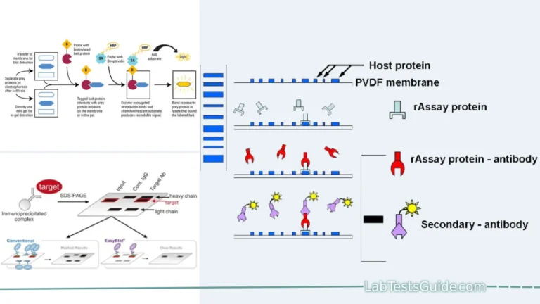
- Transfer to Membrane: After separation, the proteins are transferred from the gel to a solid support membrane, such as nitrocellulose or PVDF. This transfer is typically accomplished through electroblotting, where an electrical field helps transfer the proteins onto the membrane.
- Blocking: To prevent non-specific binding of antibodies and other detection reagents, the membrane is treated with a blocking solution, usually containing proteins like non-fat milk or bovine serum albumin.
- Primary Antibody Incubation: Specific primary antibodies that recognize the target protein(s) of interest are applied to the membrane. These antibodies bind selectively to the target protein(s).
- Washing: Unbound primary antibodies are removed through washing steps to reduce background noise.
- Secondary Antibody Incubation: A secondary antibody conjugated to an enzyme, fluorophore, or other detection moiety is applied. This secondary antibody recognizes and binds to the primary antibody, amplifying the signal.
- Washing (Again): Unbound secondary antibodies are washed away to reduce background signal.
- Detection: The presence of the target protein(s) is visualized using a suitable detection method. Common methods include chemiluminescence, fluorescence, or colorimetry, depending on the secondary antibody used.
- Analysis: The developed blot is analyzed to visualize and quantify the target protein(s) based on the intensity of bands or spots corresponding to them. Specialized software may be used for quantification.
- Control Samples: To ensure the reliability of results, control samples (e.g., positive and negative controls) are often included in the blotting experiment.
- Reference Proteins: Housekeeping proteins or loading controls are frequently used as references to normalize protein expression data.
- Reproducibility: To ensure reproducibility, experiments are often repeated and validated using statistical methods.
- Verification: Results obtained from proteomic blotting are typically verified through complementary techniques like mass spectrometry or immunoprecipitation.
- Data Interpretation: The data obtained from proteomic blotting are interpreted to draw conclusions about protein expression, post-translational modifications, or interactions.
Procedure for Proteomic Blotting:
- Sample Preparation:
- Extract proteins from the biological sample (e.g., cells or tissues).
- Denature the proteins if necessary.
- Gel Electrophoresis:
- Separate proteins based on size, charge, or both using gel electrophoresis.
- Options include one-dimensional (1D) or two-dimensional (2D) electrophoresis.
- Transfer to Membrane:
- Transfer separated proteins from the gel to a solid support membrane (e.g., nitrocellulose or PVDF).
- Employ electroblotting to transfer proteins.
- Blocking:
- Block the membrane with a blocking solution (e.g., non-fat milk) to prevent nonspecific binding.
- Primary Antibody Incubation:
- Incubate the membrane with specific primary antibodies that recognize the target protein(s).
- Washing:
- Wash the membrane to remove unbound primary antibodies.
- Secondary Antibody Incubation:
- Apply a secondary antibody conjugated to an enzyme, fluorophore, or other detection moiety.
- This secondary antibody recognizes and binds to the primary antibody.
- Washing (Again):
- Wash the membrane to remove unbound secondary antibodies.
- Detection:
- Visualize the presence of the target protein(s) using appropriate detection methods (e.g., chemiluminescence, fluorescence, or colorimetry).
- Analysis:
- Analyze the blot to quantify the target protein(s) based on band or spot intensity.
- Specialized software can be used for quantification.
- Control Samples:
- Include positive and negative controls to ensure experiment reliability.
- Reference Proteins:
- Use housekeeping proteins or loading controls as references for normalization.
- Reproducibility:
- Conduct repeat experiments to ensure reproducibility.
- Validate results using statistical methods.
- Verification:
- Verify results with complementary techniques like mass spectrometry or immunoprecipitation.
- Data Interpretation:
- Interpret data to draw conclusions about protein expression, post-translational modifications, or interactions.
Materials and Reagents:
- Polyacrylamide Gel: Used for gel electrophoresis to separate proteins.
- Transfer Membrane: Typically nitrocellulose or PVDF for protein transfer.
- Blocking Solution: Often non-fat milk or bovine serum albumin (BSA) for blocking nonspecific binding.
- Primary Antibodies: Specific antibodies recognizing target proteins.
- Secondary Antibodies: Conjugated to enzymes or fluorophores for protein detection.
- Electrophoresis Buffer: For running the gel.
- Transfer Buffer: Used during protein transfer from gel to membrane.
- Blocking Buffer: Diluent for blocking solution.
- Washing Buffer: Used for washing steps to remove unbound antibodies.
- Detection Reagents: Chemiluminescent, fluorescent, or colorimetric reagents for visualizing proteins.
- Precision Protein Standards: Molecular weight markers for size determination.
- Sample Loading Buffer: Used to prepare protein samples for gel electrophoresis.
- Protease Inhibitors: Added to lysis buffers to prevent protein degradation.
- Reducing Agent: e.g., DTT or β-mercaptoethanol, used to reduce disulfide bonds in proteins.
- Electrophoresis Equipment: Gel electrophoresis apparatus, power supply, and transfer equipment.
- Incubation Trays: For antibody incubation steps.
- Chemiluminescence Detection System: Required for chemiluminescent detection.
- Fluorescence Imaging System: Needed for fluorescence-based detection.
- Microplate Reader: For colorimetric detection methods.
- Laboratory Consumables: Pipettes, tubes, and other lab supplies.
Step-by-Step Protocol:
- Sample Preparation:
- Extract proteins from your biological sample using an appropriate lysis buffer with protease inhibitors.
- Quantify protein concentration using a protein assay like Bradford or BCA.
- Gel Electrophoresis:
- Prepare a polyacrylamide gel according to your experimental needs (e.g., percentage of the gel, sample wells).
- Load protein samples mixed with sample loading buffer onto the gel.
- Run the gel using the appropriate electrophoresis buffer until proteins are separated.
- Protein Transfer:
- Cut a piece of transfer membrane to match the gel size.
- Soak the membrane in transfer buffer.
- Assemble the transfer apparatus and transfer the separated proteins from the gel onto the membrane using electroblotting.
- Blocking:
- Incubate the membrane in blocking solution (e.g., 5% non-fat milk or BSA in a suitable buffer) to block nonspecific binding sites. Incubation time is typically 1-2 hours at room temperature or overnight at 4°C.
- Primary Antibody Incubation:
- Incubate the membrane with primary antibodies diluted in blocking buffer. The dilution should follow the antibody manufacturer’s recommendations.
- Incubate for 1-2 hours at room temperature or overnight at 4°C with gentle agitation.
- Washing:
- Wash the membrane multiple times (usually 3-5 times) with washing buffer to remove unbound primary antibodies.
- Secondary Antibody Incubation:
- Incubate the membrane with secondary antibodies conjugated to enzymes or fluorophores. The dilution should follow the manufacturer’s recommendations.
- Incubate for 1-2 hours at room temperature with gentle agitation.
- Washing (Again):
- Wash the membrane multiple times with washing buffer to remove unbound secondary antibodies.
- Detection:
- Visualize the presence of the target protein(s) using an appropriate detection method (e.g., chemiluminescence, fluorescence, or colorimetry) according to the manufacturer’s instructions.
- Analysis:
- Analyze the developed blot using suitable imaging equipment or a microplate reader, if necessary.
- Quantify the target protein(s) based on band or spot intensity using specialized software.
- Controls:
- Include positive and negative controls to validate the experiment.
- Data Interpretation:
- Interpret the results to draw conclusions about protein expression, post-translational modifications, or interactions.
Result Interpretation:
- Visual Inspection: Examine the developed blot for the presence of bands or spots that correspond to your target proteins. Bands/spots are typically darker or fluorescent in the area where the protein is detected.
- Size Determination: Determine the size of the detected protein(s) by comparing their migration distance on the blot to that of molecular weight markers (standards) run alongside the samples during gel electrophoresis.
- Quantification: Use specialized software to quantify the intensity of the bands/spots corresponding to the target proteins. This quantification provides information about relative protein abundance.
- Normalization: Normalize the protein intensities to a suitable reference, often a housekeeping protein or loading control, to account for variations in sample loading and transfer.
- Comparative Analysis: If conducting comparative proteomics, compare the protein expression levels between different samples or conditions. Look for differences or similarities in protein abundance patterns.
- Post-translational Modifications: If studying post-translational modifications, analyze shifts in protein migration patterns, which can indicate modifications such as phosphorylation or glycosylation.
- Signal Specificity: Verify the specificity of the signal by checking if the bands/spots appear at the expected molecular weight for your target protein(s).
- Positive and Negative Controls: Assess the results of your positive and negative controls to ensure the reliability of your experiment.
- Replicates and Statistics: If applicable, analyze data from replicate experiments and consider statistical tests to determine the significance of differences.
- Cross-Validation: Consider cross-validating the results with other techniques such as mass spectrometry or immunoprecipitation to confirm protein identification and interactions.
- Qualitative vs. Quantitative Data: Distinguish between qualitative data (presence/absence of a protein) and quantitative data (changes in protein abundance) in your interpretation.
- Biological Implications: Interpret the results in the context of your research question or hypothesis. Consider how the observed protein patterns relate to the biological processes you are investigating.
- Reporting: Present your results clearly in figures and tables, including molecular weights, normalized intensities, and statistical values if applicable.
- Discussion: In the discussion section of your research, provide an in-depth interpretation of the results, addressing their biological significance and relevance to your research goals.
- Literature Comparison: Compare your findings with existing literature and previous studies to contextualize your results within the broader field of research.
- Future Directions: Suggest potential future experiments or research directions based on your results and any unanswered questions that arise from your interpretation.
Troubleshooting and Tips:
- Weak Signal:
- Increase antibody concentration during incubation.
- Check the quality of antibodies and ensure they are not expired.
- High Background:
- Optimize blocking conditions to reduce nonspecific binding.
- Ensure thorough washing steps to remove unbound antibodies.
- Multiple Bands/Smearing:
- Optimize gel electrophoresis conditions, such as voltage and run time.
- Check protein integrity during sample preparation.
- No Signal:
- Verify the presence of your target protein in your sample.
- Confirm the specificity and quality of primary antibodies.
- Uneven Protein Transfer:
- Ensure even pressure during electroblotting.
- Verify the quality of transfer membranes.
- Non-uniform Gel Loading:
- Use loading controls or housekeeping proteins for normalization.
- Be consistent with sample loading volumes.
- Protein Aggregation:
- Avoid overloading the gel with protein samples.
- Use proper denaturing agents in sample preparation.
- Inconsistent Replicates:
- Pay close attention to consistency in all steps of the protocol.
- Repeat the experiment with the same conditions to confirm results.
- Low Sensitivity:
- Consider using more sensitive detection methods (e.g., enhanced chemiluminescence).
- Increase the exposure time for imaging.
- Variability Between Blots:
- Standardize your experimental conditions and reagents.
- Normalize to a stable reference protein on each blot.
- Contamination:
- Maintain a clean laboratory environment to prevent contamination.
- Use fresh buffers and reagents.
- Non-specific Antibody Binding:
- Choose high-quality antibodies with minimal cross-reactivity.
- Optimize blocking and washing steps.
- Failed Reproducibility:
- Document your protocol meticulously for future replication.
- Validate your results with complementary techniques.
- Insufficient Sample Quantity:
- Ensure you have an adequate amount of protein for detection.
- Concentrate samples if necessary.
- Experimental Design:
- Review your experimental design and consider controls and replicates to minimize errors.
- Seek Expert Advice:
- If troubleshooting doesn’t resolve issues, consult with experienced colleagues or experts in the field for guidance.
Advantages and Disadvantages of Proteomic Blotting:
Advantages:
- Specific Protein Detection: Allows the specific detection of target proteins within complex mixtures.
- Quantification: Provides quantitative information about protein expression levels.
- Versatility: Adaptable to various detection methods (chemiluminescence, fluorescence, etc.) and sample types.
- High Sensitivity: Can detect low-abundance proteins.
- Protein Profiling: Helps create protein profiles to understand complex biological systems.
- Protein-Protein Interactions: Useful for studying protein-protein interactions.
- Biomarker Discovery: Aids in identifying disease-specific biomarkers.
- Clinical Diagnostics: Used in clinical laboratories for disease diagnosis and monitoring.
- Drug Development: Crucial in drug target validation and assessing drug effects on proteins.
- Translational Research: Bridges the gap between basic research and clinical applications.
Disadvantages:
- Limited Specificity: Antibody specificity can sometimes be a limitation if antibodies cross-react with multiple proteins.
- Time-Consuming: The entire process, from sample preparation to analysis, can be time-intensive.
- Costly: Antibodies, reagents, and equipment can be expensive, particularly for comprehensive proteomic studies.
- Artifacts: Artifacts, such as non-specific bands or spots, can complicate data interpretation.
- Quantification Challenges: Quantification can be challenging and may require careful standardization.
- Limited to Known Targets: Relies on the availability of specific antibodies for known target proteins.
- Sample Complexity: Complex samples may require extensive fractionation or purification steps.
- Reproducibility: Achieving consistent and reproducible results can be challenging.
- Protein Modifications: May not detect post-translational modifications without specialized techniques.
- Cross-Reactivity: Antibodies may cross-react with similar proteins, leading to false positives.
Limitations of Proteomic Blotting:
- Specificity: Relies on the availability of specific antibodies, limiting detection to known target proteins.
- Cross-Reactivity: Antibodies may cross-react with similar proteins, leading to false positives.
- Protein Modifications: May not detect post-translational modifications without specialized techniques.
- Quantification Challenges: Quantification can be challenging and may require careful standardization.
- Sample Complexity: Complex samples may require extensive fractionation or purification steps.
- Reproducibility: Achieving consistent and reproducible results can be challenging.
- Limited to Known Targets: Limited to the study of proteins for which specific antibodies are available.
- Protein Degradation: Susceptible to degradation of proteins during sample preparation.
- Time-Consuming: The entire process, from sample preparation to analysis, can be time-intensive.
- Costly: Antibodies, reagents, and equipment can be expensive, particularly for comprehensive proteomic studies.
Variations and Modern Alternatives:
Variations:
- Two-Dimensional Gel Electrophoresis (2D-PAGE): Combines isoelectric focusing and SDS-PAGE for improved separation in proteomic analysis.
- Reverse-Phase Protein Arrays (RPPA): Allows high-throughput quantification of multiple proteins on a single chip.
- Immunoprecipitation-Western Blot (IP-Western): Combines immunoprecipitation with Western blotting to study protein-protein interactions.
- Phospho-Proteomic Blotting: Focuses on the detection of phosphorylated proteins to study signaling pathways.
- Proteomic Blotting with Mass Spectrometry (MS): Combines Western blotting with MS for protein identification and post-translational modification analysis.
Modern Alternatives:
- Mass Spectrometry-Based Proteomics: Allows comprehensive protein identification, quantification, and post-translational modification analysis.
- Next-Generation Sequencing (NGS): RNA-Seq and DNA-Seq techniques for transcriptomics and genomics research.
- Protein Microarrays: High-throughput platforms for studying protein interactions, protein profiling, and biomarker discovery.
- Single-Cell Proteomics: Enables the analysis of proteins at the single-cell level, providing insights into cellular heterogeneity.
- Cryo-Electron Microscopy (Cryo-EM): High-resolution imaging technique for studying protein structures and complexes.
- Functional Genomics: CRISPR-Cas9 and RNA interference (RNAi) for gene knockdown or knockout studies.
- Metabolomics: Analyzing small molecule metabolites for a comprehensive view of cellular processes.
Comparison of Proteomic Blotting with Modern Techniques:
| Aspect | Proteomic Blotting | Mass Spectrometry-Based Proteomics | Next-Generation Sequencing (NGS) | Protein Microarrays |
|---|---|---|---|---|
| Type of Analysis | Protein detection and quantification | Comprehensive protein identification, quantification, and PTM analysis | Transcriptomics (RNA-Seq), Genomics (DNA-Seq) | Protein interactions, profiling, and biomarker discovery |
| Detection Sensitivity | Moderate to high sensitivity for specific targets | High sensitivity for a wide range of proteins | Low sensitivity for proteins, high for nucleic acids | Moderate to high sensitivity for specific proteins |
| Sample Complexity | Suitable for complex protein mixtures | Suitable for complex protein mixtures | Suitable for DNA/RNA samples | Suitable for studying protein interactions and profiling |
| Throughput | Moderate throughput | High throughput | High throughput | High throughput |
| Detection Range | Limited to known target proteins | Broad range of proteins, including low-abundance and PTM-modified proteins | Limited to nucleic acids (genes and transcripts) | Limited to known targets |
| Quantification Accuracy | Moderate to good accuracy | Excellent accuracy, suitable for relative and absolute quantification | Good accuracy for gene expression levels | Moderate to good accuracy |
| Post-translational Modification (PTM) Analysis | Limited unless specific PTM-specific antibodies are used | Comprehensive PTM analysis, including phosphorylation, glycosylation, etc. | Limited PTM information from nucleic acid data | Limited PTM analysis |
| Protein Structure Information | Limited structural information | Limited structural information | No protein structure information | No protein structure information |
| Cellular Heterogeneity | Analysis at the population level | Can analyze heterogeneity at the single-cell level | Analyzes cellular heterogeneity at the genomic and transcriptomic levels | Limited ability to analyze cellular heterogeneity |
| Gene Expression Analysis | No gene expression data | Gene expression data (mRNA levels) can be obtained | Comprehensive gene expression data | Limited gene expression data |
| Biomarker Discovery | Limited to known biomarkers | Enables biomarker discovery for novel and known biomarkers | Limited to gene-based biomarker discovery | High-throughput biomarker discovery |
| Data Analysis Complexity | Moderate data analysis complexity | High data analysis complexity | High data analysis complexity | Moderate data analysis complexity |
| Cost | Moderate cost | High cost (expensive equipment, consumables, and expertise) | Moderate cost (depends on sequencing depth) | Moderate cost |
| Example Applications | Disease biomarker validation, protein expression analysis | Proteome-wide protein identification, PTM analysis | Gene expression profiling, mutation detection | Protein interaction studies, biomarker discovery |
| Limitations | Limited to known targets, lower sensitivity for low-abundance proteins | High cost, complexity, requires specialized equipment | Limited to nucleic acids, limited ability to detect post-translational modifications | Limited to known targets, may require custom array design |
FAQs:
1. What is proteomic blotting?
- Proteomic blotting is a laboratory technique used to detect and analyze specific proteins within complex biological samples. It involves the separation of proteins, their transfer onto a membrane, and subsequent detection using specific antibodies.
2. How does proteomic blotting differ from genomics and transcriptomics?
- Proteomic blotting focuses on studying proteins, while genomics and transcriptomics deal with genes and gene expression, respectively. Proteomics provides insights into protein levels, modifications, and interactions.
3. What is the purpose of blocking in proteomic blotting?
- Blocking is done to prevent nonspecific binding of antibodies and other detection reagents to the membrane, ensuring that they bind only to the target proteins of interest.
4. What are primary and secondary antibodies in proteomic blotting?
- Primary antibodies are specific antibodies that recognize and bind to the target proteins on the membrane. Secondary antibodies are conjugated to enzymes, fluorophores, or other detection moieties and are used to amplify the signal by binding to the primary antibodies.
5. What is the significance of loading controls in proteomic blotting?
- Loading controls, often referred to as housekeeping proteins, are used as reference proteins to normalize protein expression data, accounting for variations in sample loading and transfer.
6. What are some common troubleshooting issues in proteomic blotting?
- Common issues include weak signals, high background, multiple bands/smearing, no signal, and variations between replicates. Troubleshooting may involve adjusting antibody concentrations, optimizing electrophoresis conditions, and ensuring proper sample preparation.
7. What are the limitations of proteomic blotting?
- Limitations include the reliance on specific antibodies, potential cross-reactivity of antibodies, limited sensitivity for low-abundance proteins, and challenges in quantification and reproducibility.
8. How does proteomic blotting compare to mass spectrometry-based proteomics?
- Proteomic blotting is targeted and focuses on specific proteins, while mass spectrometry-based proteomics provides comprehensive identification and quantification of proteins, including post-translational modifications.
9. What are some modern alternatives to proteomic blotting in proteomics research?
- Modern alternatives include mass spectrometry-based proteomics, next-generation sequencing (NGS) for transcriptomics and genomics, protein microarrays, and single-cell proteomics.
10. How can I ensure the reliability of my proteomic blotting results?
- To ensure reliability, use high-quality antibodies, standardize experimental conditions, include appropriate controls, and consider validation with complementary techniques.
Conclusion:
In conclusion, proteomic blotting is a valuable laboratory technique used to detect and analyze specific proteins within complex biological samples. It plays a pivotal role in various fields of research, including molecular biology, clinical diagnostics, and drug development. Key aspects of proteomic blotting include protein separation, transfer to a membrane, antibody-based detection, and result interpretation.
While proteomic blotting has its advantages, such as specific protein detection and quantification, it also has limitations, including reliance on specific antibodies and challenges in quantification and reproducibility. Researchers often complement this technique with modern alternatives like mass spectrometry-based proteomics and next-generation sequencing to address specific research questions and overcome limitations.