Neurons (also called neurons or nerve cells) are the fundamental units of the brain and nervous system, the cells responsible for receiving sensory information from the external world, for sending motor commands to our muscles and for transforming and transmitting electrical signals at all times. . step in the middle. More than that, your interactions define who we are as people. That said, our approximately 100 billion neurons interact closely with other types of cells, broadly classified as glia (they may actually outnumber neurons, although it’s not really known).

Defination of Nerve Cells (Neurons):
Neurons, also known as nerve cells, send and receive signals from the brain. While neurons have much in common with other cell types, they are structurally and functionally unique.
Structure of Nerve Cells (Neurons):
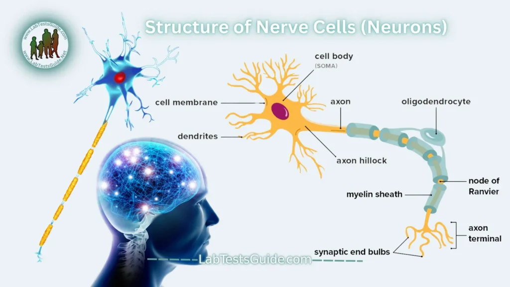
Neurons can only be seen with a microscope and can be divided into three parts:
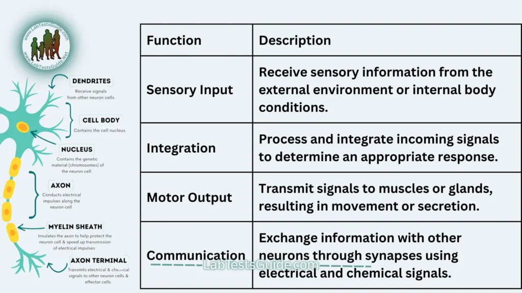
- Soma (cell body): This portion of the neuron receives information. It contains the nucleus of the cell.
- Dendrites: These thin filaments carry information from other neurons to the soma. They are the “input” part of the cell.
- Axon: This long projection carries information from the soma and sends it to other cells. This is the “output” part of the cell. It usually ends with several synapses that connect to the dendrites of other neurons.
Both dendrites and axons are sometimes called nerve fibers.
Axons vary greatly in length. Some may be tiny, while others may be over 1 meter long. The longest axon is called the dorsal root ganglion (DRG), a group of nerve cell bodies that carries information from the skin to the brain. Some of the DRG axons travel from the toes to the brain stem (up to 2 meters in a tall person).
Types of Nerve Cells (Neurons):
Neurons vary in structure, function, and genetic makeup. Given the large number of neurons, there are thousands of different types, just as there are thousands of species of living organisms on Earth.
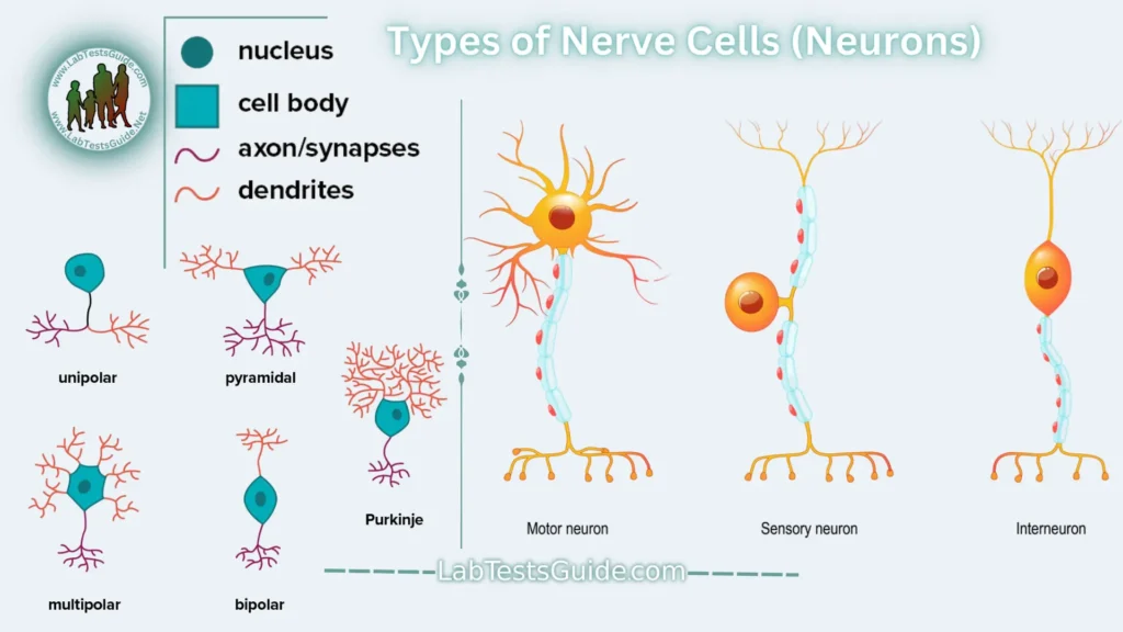
In terms of function, scientists classify neurons into three broad types: sensory, motor, and interneurons.
- Afferent Neurons (Sensory Neurons): transmit sensory information from sensory receptors (such as touch, taste, smell, vision, and hearing) to the central nervous system.
- Efferent Neurons (Motor Neurons): Located entirely within the central nervous system, they coordinate signals from sensory neurons and transmit signals to motor neurons.
- Interneurons (or Association Neurons): transmit signals from the central nervous system to muscles or glands, producing movement or secretion.
However, there are five main neuronal forms based on their morphology and locations. Each combines several elements of the basic shape of the neuron.
- Multipolar neurons. These neurons have a single axon and symmetrical dendrites extending from it. This is the most common form of neuron in the central nervous system.
- Unipolar neurons. Typically only found in invertebrate species, these neurons have a single axon.
- Bipolar neurons. Bipolar neurons have two extensions that extend from the cell body. At the end of one side is the axon and the dendrites are on the other side. These types of neurons are mainly found in the retina of the eye. But they can also be found in parts of the nervous system that help the nose and ears function.
- Pyramidal neurons. These neurons have one axon but several dendrites to form a pyramid-like shape. These are the largest neuronal cells and are mainly found in the cortex. The cortex is the part of the brain responsible for conscious thoughts.
- Purkinje neurons. Purkinje neurons have multiple dendrites that fan out from the cell body. These neurons are inhibitory neurons, meaning they release neurotransmitters that prevent other neurons from firing.
1. Censory Neurons
Sensory Neurons help you:
- Taste
- Smell
- Hear
- See
- Feel the things around you
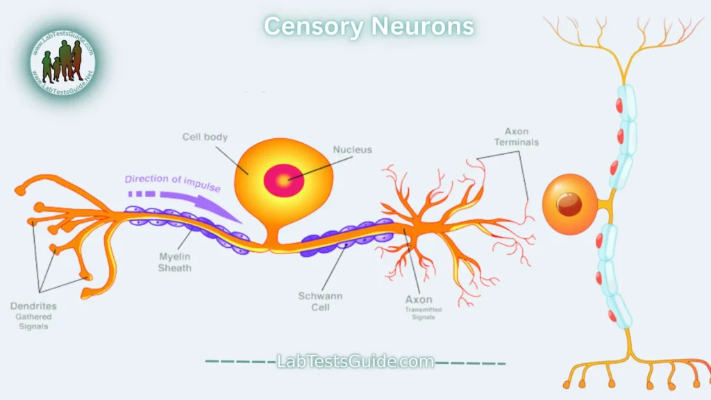
Sensory neurons are activated by physical and chemical inputs from the environment. Sound, touch, heat and light are physical inputs. Smell and taste are chemical inputs.
For example, stepping on hot sand activates sensory neurons in the soles of your feet. Those neurons send a message to your brain, making you aware of the heat.
2. Motor Neurons:
Motor neurons play a role in movement, including voluntary and involuntary movements. These neurons allow the brain and spinal cord to communicate with muscles, organs, and glands throughout the body.
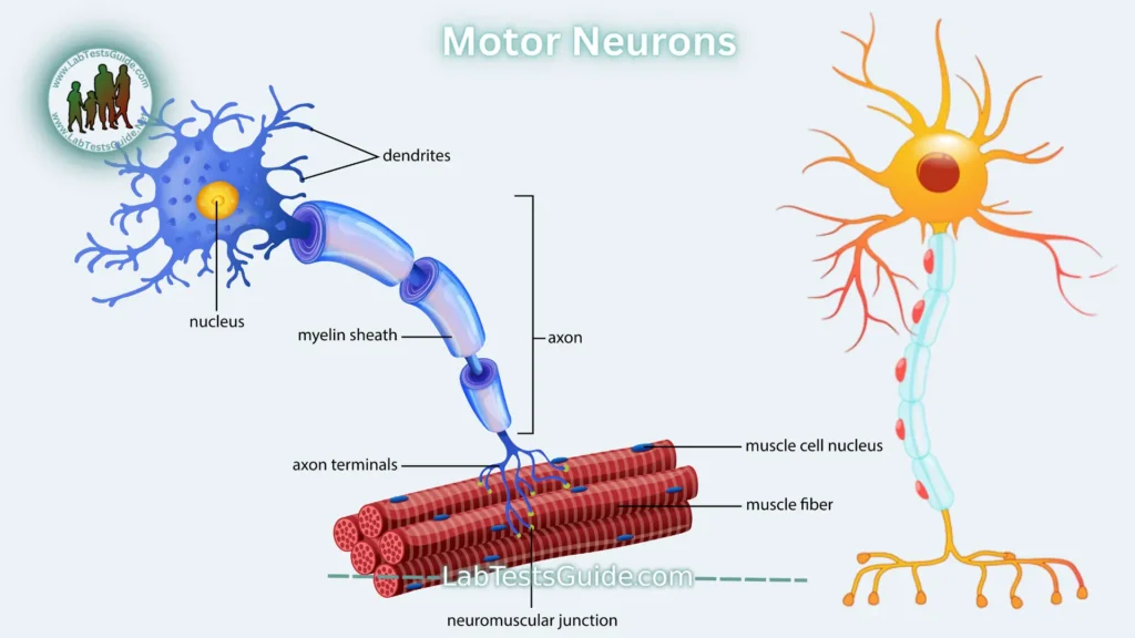
There are two types of motor neurons: lower and upper. Lower motor neurons carry signals from the spinal cord to smooth and skeletal muscles. Upper motor neurons carry signals between the brain and spinal cord.
When you eat, for example, lower motor neurons in the spinal cord send signals to the smooth muscles of the esophagus, stomach, and intestines. These muscles contract, allowing food to move through the digestive tract.
3. Interneurons (or Association Neurons):
Interneurons are neuronal intermediates found in the brain and spinal cord. They are the most common type of neuron. They pass signals from sensory neurons and other interneurons to motor neurons and other interneurons. They often form complex circuits that help you react to external stimuli.
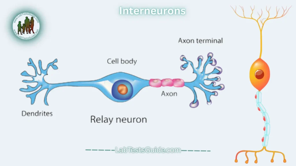
For example, when you touch something sharp like a cactus, sensory neurons in your fingertips send a signal to interneurons in your spinal cord. Some interneurons transmit the signal to the motor neurons in the hand, allowing you to move it away. Other interneurons send a signal to the pain center in your brain and you experience pain.
Functions of Nerve Cells (Neurons):
Neurons send signals using action potentials. An action potential is a change in the electrical potential of a neuron caused by the flow of charged particles in and out of the neuron’s membrane. When an action potential is generated, it is carried along the axon to a presynaptic terminal.
Action potentials can activate both chemical and electrical synapses. Synapses are places where neurons can transmit these electrical and chemical messages between themselves. Synapses consist of a presynaptic end, a synaptic cleft, and a postsynaptic end.
Chemical Synapses:
At a chemical synapse, the neuron releases chemical messengers called neurotransmitters. These molecules cross the synaptic cleft and bind to receptors at the postsynaptic end of a dendrite.
Neurotransmitters can trigger a response in the postsynaptic neuron, causing it to generate its own action potential. Alternatively, they may prevent activity in the postsynaptic neuron. In that case, the postsynaptic neuron does not generate an action potential.
Electrical Synapses:
Electrical synapses can only excite. These synapses are formed when two neurons are connected by a gap junction. This gap is much smaller than a chemical synapse and is made up of ion channels that help transmit a positive electrical signal.
Because of the way these signals travel, signals move much faster through electrical synapses than through chemical synapses. However, these signals can decrease from one neuron to the next. This makes them less effective at transmitting repeated signals.
Importance of Nerve Cells (Neurons):
Neurons play a crucial role in all aspects of nervous system function, including:
- Sensory perception: Neurons allow us to sense the world around us and to experience our own bodies.
- Movement: Neurons control our muscles, allowing us to move our bodies in response to our environment and our thoughts.
- Thinking and learning: Neurons are involved in all aspects of cognitive function, including memory, language, and emotion.
In Clinical Laboratories:
In clinical laboratories, the study of nerve cells (neurons) can be crucial in diagnosing and understanding various neurological disorders and conditions. This is how nerve cells are relevant in clinical laboratory settings:
Histopathology: In cases of neurological diseases or injuries, such as neurodegenerative disorders (e.g., Alzheimer’s disease, Parkinson’s disease) or traumatic brain injuries, pathologists may obtain tissue samples (biopsies) and examine them under a microscope. This allows evaluation of neuronal structure, the presence of abnormalities (such as neurofibrillary tangles or Lewy bodies), and overall tissue integrity.
Cytology: Cerebrospinal fluid (CSF) analysis can provide valuable information about neurological conditions. CSF contains cells removed from the central nervous system, including neurons. Abnormalities in neuron morphology or the presence of atypical cells may indicate neurological disorders such as meningitis, encephalitis, or certain types of cancer (e.g., glioblastoma).
Electrophysiology: Electrophysiological tests, such as electroencephalography (EEG) and nerve conduction studies, measure the electrical activity of neurons. These tests can help in the diagnosis of conditions that affect nerve function, such as epilepsy, neuropathies, and demyelinating diseases (e.g., multiple sclerosis).
Molecular testing: Molecular techniques, including polymerase chain reaction (PCR), next-generation sequencing (NGS), and fluorescence in situ hybridization (FISH), can be used to detect genetic mutations associated with neurological disorders. For example, genetic testing can identify mutations in genes related to neurodevelopmental disorders (for example, autism spectrum disorders) or inherited neuropathies.
Immunohistochemistry: Immunohistochemical staining of tissue samples can help identify specific neuronal markers or proteins associated with certain neurological conditions. For example, staining for beta-amyloid and tau proteins can help in the diagnosis of Alzheimer’s disease, while staining for synaptophysin can help identify neuroendocrine tumors.
Neuropathology: Neuropathologists specialize in the examination of tissues of the nervous system and play a critical role in the diagnosis of neurological diseases. They use a combination of histological, immunohistochemical and molecular techniques to characterize neuronal abnormalities and provide information on disease mechanisms.
FAQs:
What are nerve cells?
Nerve cells, or neurons, are specialized cells in the nervous system responsible for transmitting electrical and chemical signals throughout the body. They play a fundamental role in sensory perception, motor coordination, cognition, and behavior.
What is the structure of a nerve cell?
Nerve cells consist of a cell body (soma), dendrites, and an axon. The cell body contains the nucleus and other organelles necessary for cellular function. Dendrites are branch-like extensions that receive signals from other neurons, while the axon is a long, slender projection that transmits signals away from the cell body.
How do nerve cells transmit signals?
Nerve cells transmit signals through electrical impulses known as action potentials. These impulses travel along the axon and are transmitted to other neurons or effector cells (such as muscles or glands) at specialized junctions called synapses. At the synapse, neurotransmitters are released from the axon terminal and received by the dendrites or cell body of the neighboring neuron.
What are the different types of nerve cells?
There are several types of nerve cells, including sensory neurons, motor neurons, and interneurons. Sensory neurons transmit sensory information from sensory receptors to the central nervous system. Motor neurons transmit signals from the central nervous system to muscles or glands. Interneurons facilitate communication between neurons in the central nervous system.
How do nerve cells communicate with each other?
Nerve cells communicate with each other at synapses, where neurotransmitters are released from the axon terminal of one neuron and received by the dendrites or cell body of another neuron. This chemical signaling process allows for the transmission and integration of information in the nervous system.
What is the role of nerve cells in the brain?
Nerve cells in the brain are involved in various functions, including sensory processing, motor control, memory, learning, and emotional regulation. They form complex neural networks and circuits that underlie cognitive processes and behavior.
What happens if nerve cells are damaged or dysfunctional?
Damage or dysfunction of nerve cells can lead to neurological disorders and impairments in sensory, motor, or cognitive function. Neurological disorders such as Alzheimer’s disease, Parkinson’s disease, epilepsy, and stroke can result from abnormalities in nerve cell structure or function.
Can nerve cells regenerate?
In some cases, nerve cells can regenerate, particularly in the peripheral nervous system. However, regeneration in the central nervous system is more limited due to factors such as inhibitory molecules and scar formation. Research into techniques to promote nerve cell regeneration is ongoing and holds promise for the treatment of neurological injuries and disorders.
How can we protect nerve cells and maintain brain health?
Adopting a healthy lifestyle, including regular exercise, a balanced diet, adequate sleep, and cognitive stimulation, can help protect nerve cells and maintain brain health. Avoiding risk factors such as smoking, excessive alcohol consumption, and head injuries is also important for preserving neurological function.
Possible References Used


