Electrochemiluminescence Immunoassay (ECLIA)
The pinnacle of voltage-triggered diagnostic technology. Discover the unique biochemical physics of Ruthenium, the Streptavidin-Biotin system, and the revolutionary signal amplification cycle.
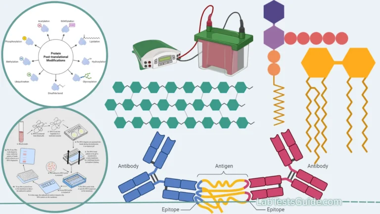
")
Introduction to Electrochemiluminescence
Definition: Electrochemiluminescence Immunoassay (ECLIA) is a highly advanced, proprietary quantitative laboratory technique that detects biological analytes through light emission. Unlike standard CLIA which uses a chemical trigger, ECLIA relies on a specific electrical voltage applied to a platinum electrode to trigger the luminescent reaction.
ECLIA (predominantly championed by Roche Diagnostics via their Elecsys and cobas platforms) represents a profound evolution in immunochemistry. By utilizing a unique metallic complex—Ruthenium—and Tripropylamine (TPA), ECLIA creates a cyclical, regenerating reaction. This means the signal amplifies itself continuously while voltage is applied, leading to an extraordinarily broad dynamic measuring range and unmatched analytical sensitivity.
This technology is currently the global standard for high-throughput testing in endocrinology, cardiac markers, infectious disease serology, and oncology, allowing labs to measure everything from highly concentrated tumor markers down to trace amounts of vitamins in a single analyzer format.
The Four Pillars of an ECLIA System
The ECLIA mechanism requires four highly specific components to execute its voltage-driven assay:
1. Biotinylated Capture Antibody
Instead of the capture antibody being permanently fixed to a bead, it floats freely in the reagent, attached to a Biotin molecule. This allows for faster, liquid-phase reaction kinetics with the patient sample.
2. Ruthenium-Labeled Conjugate
The detection antibody is labeled with a Ruthenium complex: Ru(bpy)₃²⁺. This metallic compound is the source of the luminescent signal, but it only emits light when electrically stimulated.
3. Streptavidin Microparticles
Paramagnetic beads coated in Streptavidin are added later in the incubation. Because Streptavidin binds to Biotin with one of the strongest non-covalent affinities in nature, it easily pulls the entire immune complex onto the solid phase.
4. TPA & The Measuring Cell
Tripropylamine (TPA) is a chemical added just before reading. The complex is pulled onto a working electrode by a magnet. When voltage is applied, the Ruthenium and TPA react to produce light.
The Principle of the Test: The ECLIA Cycle
The defining scientific advantage of ECLIA over standard CLIA is that the light-emitting label is not destroyed during the reaction. It is regenerated in a continuous cycle as long as voltage is applied, leading to massive signal amplification.
Step 1: Oxidation at the Electrode
A specific electrical voltage is applied to the platinum working electrode. This simultaneously oxidizes the Ruthenium complex (Ru²⁺ becomes Ru³⁺) and oxidizes the TPA molecule into a highly reactive TPA cation radical.
Step 2: Excitation & Emission
The TPA radical violently reacts with the Ru³⁺, forcing the Ruthenium into an unstable, excited state (Ru²⁺*). As the Ruthenium instantly decays back to its stable ground state, it emits a photon of light at exactly 620 nm.
Step 3: Continuous Regeneration
Because the Ruthenium has returned to its normal Ru²⁺ state, the electrode immediately oxidizes it again. A single Ruthenium label can go through this emission cycle thousands of times per second.
Advantages & Disadvantages of ECLIA
✅ Advantages
- High Sensitivity & Specificity: Capable of detecting minute picogram concentrations.
- Extremely Wide Measuring Range: The regenerative cycle allows linearity up to 6 orders of magnitude, vastly reducing the need for sample dilution.
- Rapid Turnaround Time: Standard tests take 18 minutes. STAT applications (like Troponin) yield results in exactly 9 minutes.
- Small Sample Volume: Requires very little serum/plasma (typically only 10 to 50 µL per test).
- High Reagent Stability: Ruthenium labels are incredibly stable, providing long onboard shelf life for reagents.
❌ Disadvantages & Limitations
- High Cost: Analyzers and proprietary reagents are expensive to acquire and maintain.
- Closed System: Reagents are entirely proprietary (predominantly manufactured by Roche).
- Biotin Interference: The streptavidin-biotin solid phase is highly vulnerable to exogenous Biotin supplements.
- Hardware Maintenance: The platinum flow cell electrode must be meticulously cleaned (CleanCell process) after every single test to prevent protein carryover.
Comparison: ECLIA vs. CLIA vs. ELISA
Understanding how ECLIA differs from standard luminescent and colorimetric assays highlights its clinical superiority.
| Feature | ECLIA (Electrochemiluminescence) | CLIA (Standard Chemiluminescence) | ELISA (Enzyme-Linked) |
|---|---|---|---|
| Trigger Mechanism | Electrical Voltage + TPA | Chemical Trigger (Acid/Base) | Enzyme + Substrate |
| Label Type | Ruthenium Ru(bpy)₃²⁺ | Acridinium Ester / Isoluminol | Enzymes (HRP, ALP) |
| Signal Generation | Cyclical / Regenerating Light | Single Flash / Glow | Color Change (Absorbance) |
| Test Duration | 9 to 18 Minutes | 15 to 45 Minutes | 1 to 3 Hours |
| Interference Risk | High (Biotin, Fibrin on electrode) | Moderate (Biotin, HAMA) | Moderate (Turbidity, Color) |
Pre-Analytical Parameters & Specimen Collection
Because ECLIA relies fundamentally on the Streptavidin-Biotin capture system, it is highly susceptible to exogenous Biotin interference. Patients consuming high doses of Biotin (Vitamin B7, >5 mg/day)—often found in hair/skin/nail supplements or prescribed for Multiple Sclerosis—will have excess free biotin in their blood.
This free biotin binds to the Streptavidin beads first, blocking the assay’s immune complex from attaching. This causes falsely low results in Sandwich assays (like TSH, Troponin) and falsely high results in Competitive assays (like Free T4, Vitamin D). Patients must cease biotin supplements 12–72 hours prior to the blood draw.
Sample Requirements
- Specimen: Serum (SST) is standard. Lithium Heparin or EDTA plasma is acceptable for most, but not all, assays.
- Storage: Highly stable; usually 7 days at 2°C–8°C. Can be frozen at -20°C for longer storage (thaw thoroughly and vortex before use).
- Centrifugation: Must be thoroughly centrifuged. Fibrin strands or RBCs can coat the platinum electrode, physically blocking the voltage and causing total assay failure.
Assay Formats Used in ECLIA
- Sandwich Assay: Used for large, multi-epitope molecules (TSH, PSA, FSH, Troponin). The signal is directly proportional to analyte concentration.
- Competitive Assay: Used for small molecules (Free T3, Vitamin D, Testosterone). Unlabeled patient antigen competes with Ruthenium-labeled antigen. The signal is inversely proportional to analyte concentration.
The Automated ECLIA Protocol Timeline
This timeline represents the exact sequence executed inside the measuring cell of an ECLIA analyzer (such as the Roche cobas e411, e601, or e801).
1. First Incubation (Liquid Phase)
The patient sample is mixed with the Biotinylated Capture Antibody and the Ruthenium-Labeled Detection Antibody. Because there are no heavy beads yet, the reaction happens rapidly in a liquid phase at exactly 37°C.
2. Second Incubation (Solid Phase Binding)
Streptavidin-coated paramagnetic microparticles are added. The Biotin (attached to the now-formed immune complex) binds irreversibly to the Streptavidin, anchoring the complex to the magnetic beads.
3. Aspiration into the Measuring Cell
The reaction mixture is sucked into the flow cell. A powerful magnet positioned directly beneath the working electrode activates, pulling the magnetic beads tightly onto the surface of the platinum electrode.
4. Wash & ProCell (TPA) Addition
ProCell buffer fluid flows over the electrode, washing away all unbound serum proteins and excess reagents. Simultaneously, this fluid fills the cell with the necessary Tripropylamine (TPA) co-reactant.
5. Voltage Trigger & PMT Reading
Voltage is applied to the electrode. The Ruthenium/TPA light emission cycle begins instantly. A Photomultiplier Tube (PMT) located directly above the electrode captures the photons, converting them into a digital signal.
6. CleanCell Process
The magnet turns off, and a highly specific cleaning solution (CleanCell) washes the beads away and chemically conditions the platinum electrode so it is pristine for the very next patient test.
Optical Signal Diagnostics: ECLIA S/CO Analysis
In qualitative ECLIA systems (like Hepatitis or HIV screening), the PMT reads the light signal and calculates a Cut-Off Index (COI) or S/CO ratio. This tool simulates how the analyzer flags results.
Primary Clinical Applications of ECLIA
Because of its incredible detection range and speed, ECLIA is heavily utilized for advanced clinical chemistry testing across several medical specialties:
Endocrinology & Thyroid Function
ECLIA is famous for its Thyroid testing capabilities (TSH, FT3, FT4). A 3rd Generation TSH ECLIA assay can accurately detect concentrations as low as 0.005 µIU/mL, making it vital for diagnosing subclinical hyperthyroidism.
Cardiac Markers & Emergency
ECLIA plays a critical role in acute myocardial infarction diagnostics. High-Sensitivity Troponin T (hs-TnT) assays, utilizing 9-minute STAT protocols, can detect infinitesimal cardiac damage within 1 hour of chest pain onset. Also used for NT-proBNP and CK-MB.
Oncology (Tumor Markers)
The massive dynamic measuring range of ECLIA reduces the need to manually dilute highly concentrated tumor markers. It is heavily utilized for Total PSA, Free PSA, CEA, CA-125, AFP, and CA 19-9 therapy monitoring.
Infectious Disease Serology
Offers extreme specificity for blood banking and STD screening. Commonly runs Hepatitis panels (HBsAg, Anti-HCV), HIV 1/2 Ag/Ab Combo (4th Gen), Syphilis, and SARS-CoV-2 antibodies with minimal false positive rates.
Bone Markers, Vitamins & Anemia
Competitively formatted ECLIA is ideal for quantifying trace vitamins and metabolic markers, including 25-OH Vitamin D, Intact PTH, Vitamin B12, Folate, Ferritin, and Osteocalcin.
Fertility & Reproductive Hormones
Routinely measures reproductive health indicators like Beta-hCG (for pregnancy and certain tumors), FSH, LH, Prolactin, Estradiol, Testosterone, Progesterone, and Anti-Mullerian Hormone (AMH).
Frequently Asked Questions & Glossary
Glossary of ECLIA Terms
Ruthenium Ru(bpy)₃²⁺: The metallic complex used as the luminescent label in ECLIA.
TPA (Tripropylamine): The chemical co-reactant that facilitates the excitation of Ruthenium when voltage is applied.
Streptavidin: A protein with an extraordinarily high affinity for Biotin, used to coat the magnetic particles.
Working Electrode: A small platinum plate inside the analyzer’s measuring cell where the magnetic beads are captured and voltage is applied.
What is ECLIA?
ECLIA stands for “Electrochemiluminescence immunoassay.” It is a highly sensitive laboratory technique used to detect and quantify substances in the blood or body fluids, widely employed in medical diagnostics.
How does ECLIA work?
ECLIA involves the use of specific antibodies that selectively bind to the target analyte in the patient’s sample. A detection antibody labeled with a luminescent marker is then introduced. When an electrical current is applied, the labeled antibody undergoes an electrochemical reaction, emitting light (chemiluminescence) that is proportional to the analyte’s concentration.
What are the advantages of ECLIA over other immunoassay methods?
ECLIA offers advantages such as high sensitivity, wide dynamic range, low interference, fast results, minimal sample volume, and high automation and throughput capabilities. These features make it a preferred method in clinical diagnostics.
What is the main difference between CLIA and ECLIA?
While both produce light to quantify analytes, CLIA uses a one-time chemical trigger (like hydrogen peroxide) which destroys the label after flashing. ECLIA uses an electrical voltage trigger that constantly regenerates the Ruthenium label, creating a highly stable, continuous signal that can be tightly controlled by simply turning the voltage on or off.
Why is the “CleanCell” step so critical in ECLIA?
Unlike standard CLIA which throws away the plastic reaction cuvette after every test, ECLIA uses a permanent flow cell and platinum electrode built into the machine. If proteins or fibrin from a patient sample coat the electrode, it acts as an insulator, preventing the voltage from reaching the next patient’s sample. The CleanCell solution strips the electrode bare between every single test to prevent carryover.
How can a lab mitigate the Biotin interference issue?
Patient education is the best defense; patients should stop taking high-dose biotin 12–72 hours before a blood draw. In the lab, suspected samples can be treated with specialized “Biotin-blocking” reagents or diluents before running the assay to neutralize the free biotin, or the lab can switch the sample to an analyzer that does not use a streptavidin-biotin solid phase.
What are the limitations of ECLIA?
ECLIA limitations include potential cross-reactivity, sample interference from certain matrices, and the need for skilled operators and specialized equipment. Hemolysis or other sample issues may also impact results.
What analytes can be measured using ECLIA?
ECLIA can measure a wide range of analytes, including hormones, tumor markers, cardiac biomarkers, infectious agents, autoimmune markers, drugs, and more. Its versatility allows for various diagnostic applications.
Is ECLIA suitable for point-of-care testing (POCT)?
ECLIA is less suitable for POCT due to its requirement for specialized equipment and infrastructure. Other rapid immunoassay methods may be more appropriate for point-of-care applications.
How are ECLIA results interpreted?
ECLIA results are interpreted by comparing the obtained value with established reference ranges or cut-off values. The clinical context, patient history, and other diagnostic findings are also considered for accurate interpretation.
What are the future trends in ECLIA technology?
Future trends in ECLIA technology include enhanced sensitivity, multiplex assays, miniaturization for POCT, high-throughput automation, integration with other diagnostic methods, and personalized medicine applications.
What role does ECLIA play in medical diagnostics?
ECLIA plays a crucial role in medical diagnostics by providing accurate and precise measurements of various analytes in patient samples. It aids in the early detection, diagnosis, monitoring, and management of numerous diseases and conditions, contributing to improved patient care.