Biological Coloration: The Comprehensive Clinical & Scientific Guide
A Multi-Disciplinary Analysis of Biological Pigments, Microbial Identification, Structural Morphology, and Laboratory Diagnostic Relevance.
Introduction to Biological Coloration
According to the Oxford Dictionary, “Color” is a property of matter that allows it to absorb light of specific wavelengths and reflect others. Biological coloration is the multifaceted appearance of an organism resulting from light’s interaction with biological tissues. This phenomenon is governed by selective absorption, reflection, and emission. Every living organism varies by overall body colors and distribution patterns (color patterns).
Evolutionary Drivers
Organisms utilize traits for four primary biological drivers:
- Survival: Background matching (Camouflage) and evading detection.
- Communication: Sexual attraction, mate selection, and species recognition.
- Protection: Physical shielding from ionizing UV radiation and warning signals (Aposematism).
- Adaptation: Thermoregulation, metabolic optimization, and Mimicry.
The Coloration Spectrum
Biochromes: True chemical pigments that absorb specific light wavelengths via conjugated double bonds.
Schemochromes: Physical structures producing color through scattering, interference, or diffraction.
Chromatophores: Specialized cells such as melanophores (black), xanthophores (yellow), erythrophores (red), and iridophores (iridescence).
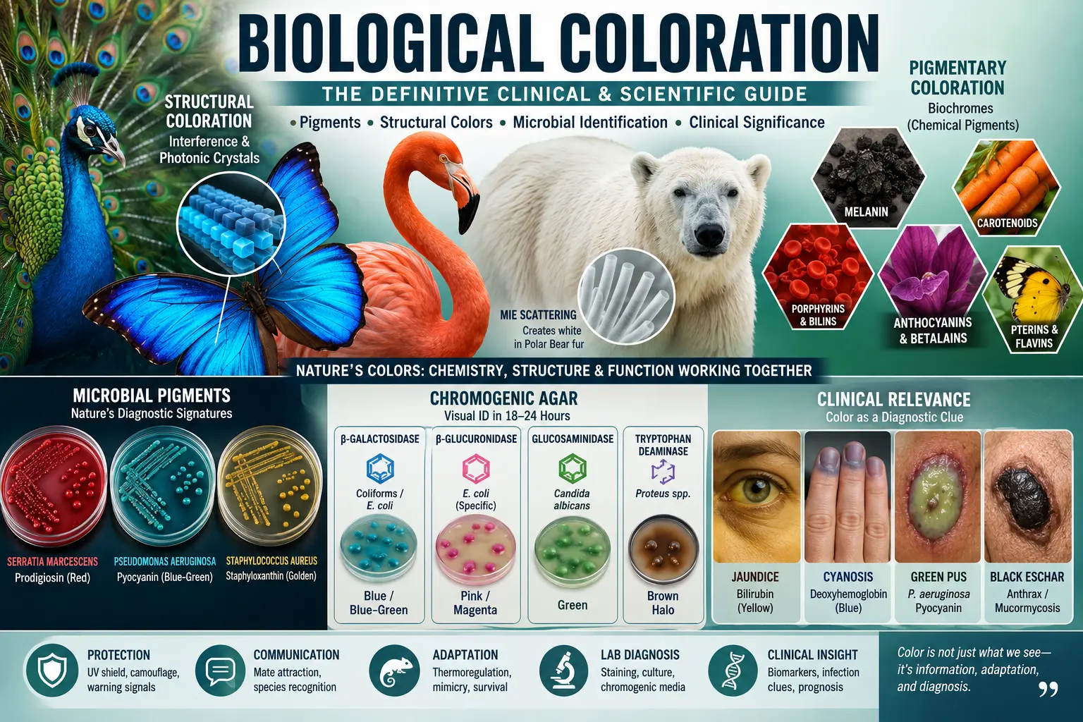
Major Pigment Families (Biochromes)
Pigments contain chromophores that selectively absorb light. Their expression is influenced by enzymes, genetics, diet, and environment.
1. Melanin Group
Produced by melanocytes from the amino acid Tyrosine via the enzyme Tyrosinase (Raper-Mason pathway).
- Eumelanin: Black and brown hues; essential for photoprotection and Industrial Melanism (e.g., Peppered Moth).
- Pheomelanin: Contains sulfur; produces yellow to reddish hues (common in red hair).
- Neuromelanin: Found in the brain’s substantia nigra; derived from dopamine.
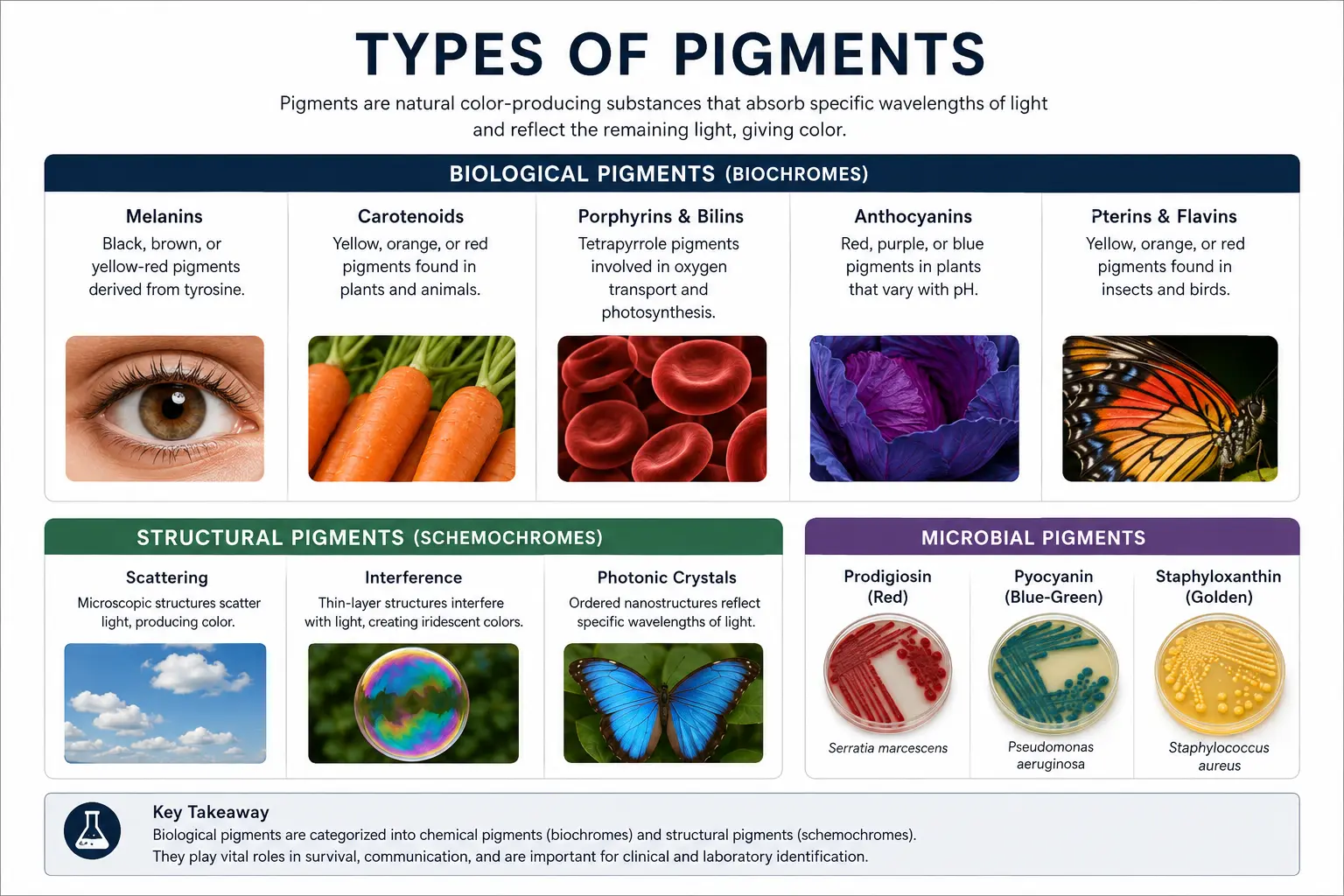
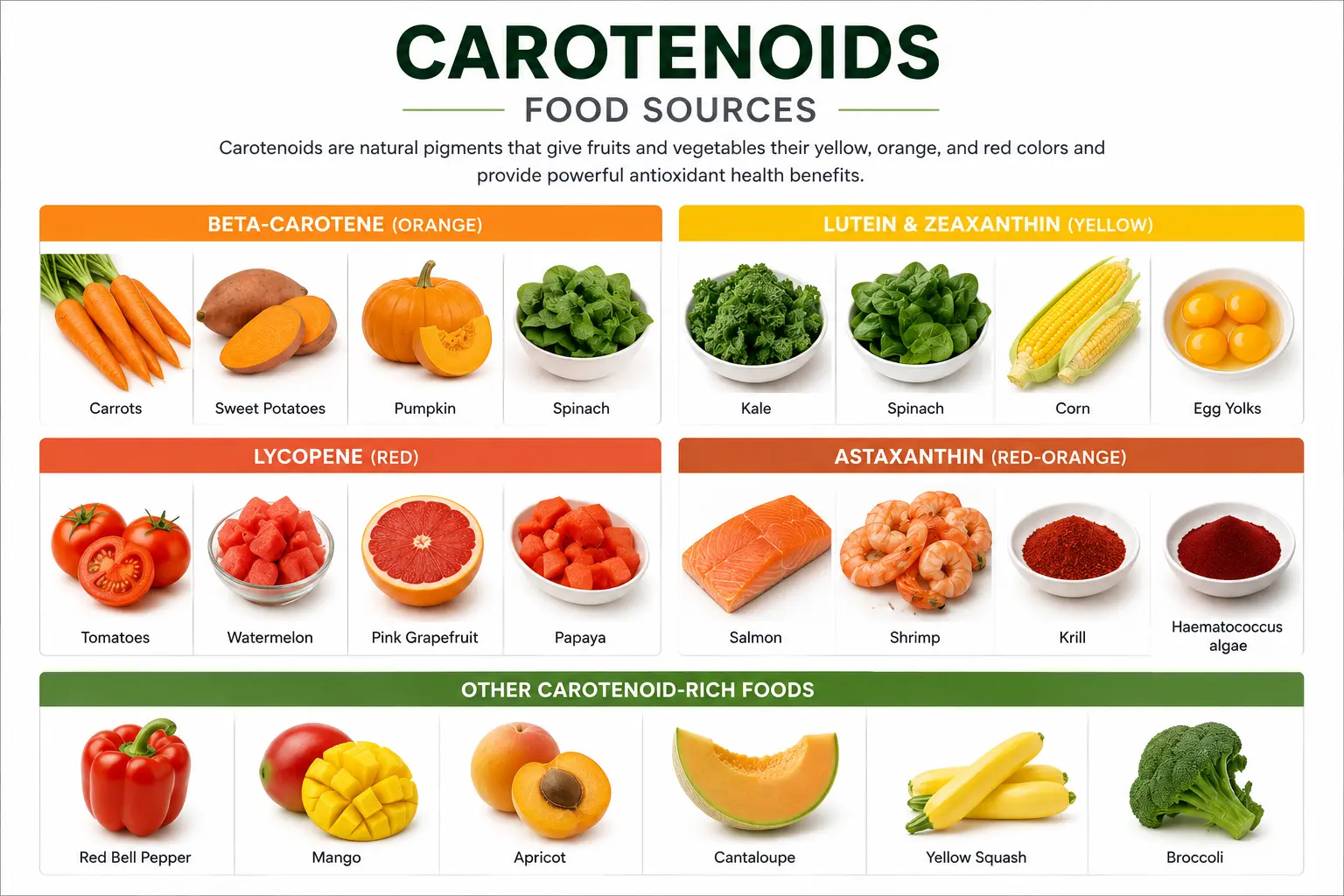
2. Carotenoids & Lipochromes
Non-nitrogenous tetraterpenes consisting of eight isoprene units. Animals must obtain these via diet.
- Carotenes: Oxygen-free. β-carotene (orange), Lycopene (red), α-carotene.
- Xanthophylls: Oxygenated. Lutein (yellow plumage), Astaxanthin (red/purple in Salmon and Flamingos), Zeaxanthin.
3. Porphyrins & Bilins
Nitrogenous tetrapyrrole rings vital for oxygen transport and photosynthesis.
- Hemoglobin: Contains Iron (Fe). Bright Red (Oxy), Maroon (Deoxy).
- Chlorophyll (a-e): Contains Magnesium (Mg). Green hue for photosynthesis.
- Bilins: Biliverdin (green) and Bilirubin (yellow) from heme breakdown.
- Ooporphyrins: Provides red/brown flecks on eggshells.
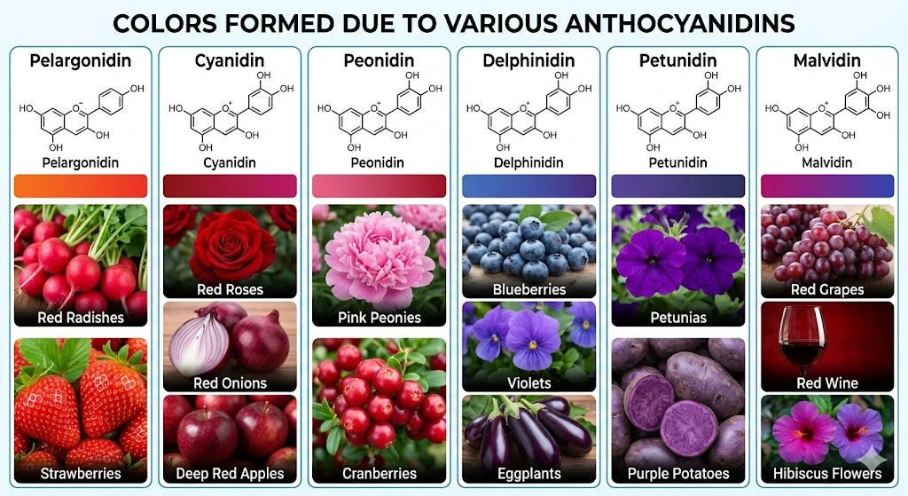
4. Anthocyanins & Betalains
- Anthocyanins: Water-soluble flavonoids (Cyanidin, Delphinidin). Color shifts with vacuolar pH (Red, Purple, Blue). Protect plants from UV stress.
- Betalains: Exclusive to Caryophyllales (Beetroot, Cactus) and fungi; replace anthocyanins.
5. Pterins & Flavins
Nitrogenous heterocyclic pigments producing red, orange, and yellow. Found in butterfly wings, grasshoppers, and some bird feathers (Xanthopterin, Drosopterin).
Structural Coloration (Schemochromes)
Colors arising from nano-sized structures interacting with light. Unlike pigments, these are often iridescent and angle-dependent.
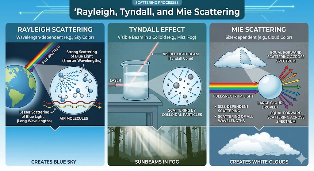
Scattering Mechanisms
Rayleigh Scattering: Microscopic structures create the blue in bird feathers.
Tyndall effect: Creates blue eye color in humans via colloidal suspension.
Mie Scattering: Hollow transparent keratin scatters all wavelengths equally, creating the White fur of Polar Bears and preventing UV penetration.
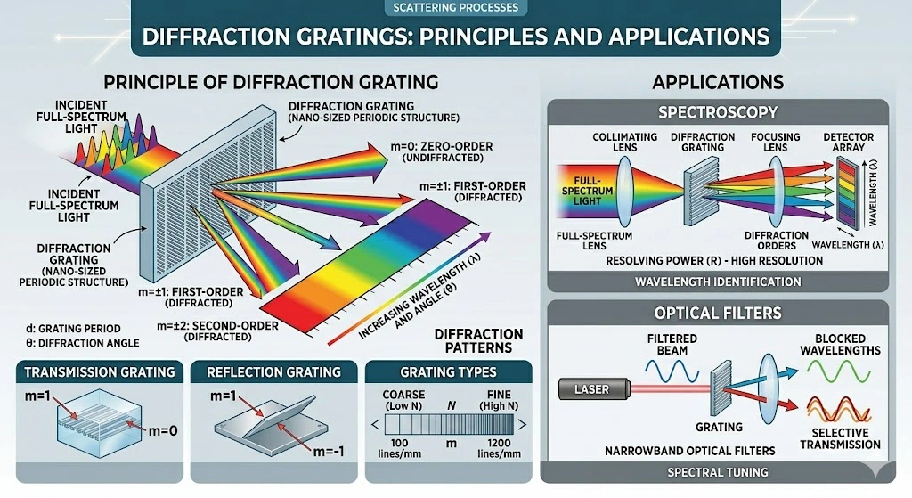
Interference & Diffraction
Thin-film Interference: Light reflects off upper/lower boundaries (Soap bubbles, Morpho butterfly wings, Oil slicks, Golden beetles).
Diffraction Gratings: Regularly spaced ridges (Peacock tail feathers, Jewel beetles) split light into a prism effect.
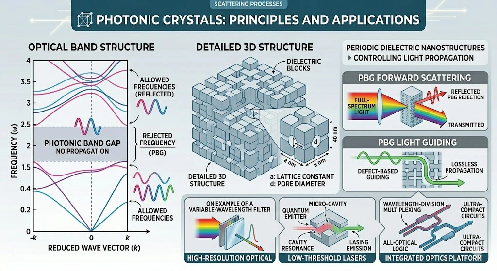
Photonic Crystals
Periodic nanostructures causing Bragg’s Reflection. Example: Pollia condensata (Marble Berry) – the most intense, fadeless blue in nature. Also found in the Emerald-patched cattle heart butterfly.
Comparison Matrix: Structural vs Pigment vs Bioluminescence
| Feature | Pigmentary (Chemical) | Structural (Physical) | Bioluminescence |
|---|---|---|---|
| Primary Mechanism | Selective Light Absorption | Reflection/Scattering/Diffraction | Chemical Reaction (Luciferase) |
| Origin | Biochromes (Melanin, Carotenoids) | Nano-structures (Photonic crystals) | Metabolic enzyme reaction |
| Angle Dependency | Static (Same from all angles) | Dynamic (Iridescent/Angle-shifting) | Active Light Emission |
| Longevity | May fade (Photobleaching) | Permanent unless structure damaged | Temporary/Active |
| Lab Relevance | Gram Stain, Chromogenic media | SEM, Biomimetics | Bio-assays and Gene markers |
Microbial Pigmentation & Lab Identification
Bacterial pigments are secondary metabolites that serve as definitive taxonomic markers and virulence factors.
Prodigiosin (Red)
Organism: Serratia marcescens
Tripyrrole pigment. Temperature sensitive: Deep red at 25°C, colorless at 37°C.
Pyocyanin (Blue-Green)
Organism: Pseudomonas aeruginosa
Redox-active phenazine. Water-soluble; diffuses into Mueller-Hinton Agar or King’s Media.
Staphyloxanthin (Golden)
Organism: Staphylococcus aureus
Carotenoid virulence factor that neutralizes host reactive oxygen species (ROS).
Chromogenic Agar Test Principles
Colorless substrates (chromogens) cleaved by specific bacterial enzymes release colored products, allowing visual identification within 18-24h.
| Enzyme Target | Organism Detected | Colony Color Result |
|---|---|---|
| β-galactosidase | Coliforms / E. coli | Blue / Blue-Green |
| β-glucuronidase | E. coli (Specific) | Pink / Magenta |
| Glucosaminidase | Candida albicans | Green |
| Tryptophan Deaminase | Proteus spp. | Brown Halo |
Biochemical Pathways & Clinical Correlation
Pathways
Melanin (Raper-Mason): Tyrosine → DOPA → Dopachrome → Melanin.
Heme: Tetrapyrrole ring with Iron (Fe²⁺).
Chlorophyll: Tetrapyrrole ring with Magnesium (Mg²⁺).
Clinical Biomarkers
Jaundice (Icterus): Yellow skin from Bilirubin elevation.
Cyanosis: Blue skin from deoxyhemoglobin hypoxia.
Melanoma: Malignancy of melanocytes.
Infection Indicators
Green Pus: P. aeruginosa pyocyanin.
Golden Crust: S. aureus Impetigo.
Black Eschar: Cutaneous Anthrax or Mucormycosis.
Exam Essentials & High-Yield Points
1. Which mechanism produces the white color in Polar Bears?
Answer: Mie Scattering (within hollow keratin fur cores).
2. What is the precursor for Melanin?
Answer: The amino acid Tyrosine.
3. Why does Serratia marcescens lose its red color at 37°C?
Answer: Prodigiosin production is temperature-dependent and inhibited at human body temperature.
4. Why do Gram-negative bacteria appear pink?
Answer: They lose the primary purple stain during decolorization and take up the Safranin counterstain.
5. What is the structural difference between Carotenes and Xanthophylls?
Answer: Carotenes are pure hydrocarbons; Xanthophylls contain oxygen.
6. What causes the blue color in human eyes?
Answer: Tyndall/Rayleigh scattering (Structural), not blue pigment.
7. Name the red pigment in Caryophyllales that replaces anthocyanin.
Answer: Betalains.
8. Which fruit features fadeless blue via Photonic Crystals?
Answer: Pollia condensata (Marble Berry).
Frequently Asked Questions
Summary & Conclusions
Biological coloration is a sophisticated interplay of biochromes (chemical: melanin, carotenoids, porphyrins, anthocyanins, betalains) and schemochromes (physical: scattering, interference, photonic crystals). From the photoprotective role of melanin and the diagnostic utility of Chromogenic Agar to adaptive strategies like Aposematism and Industrial Melanism, coloration is a vital tool for survival, communication, and clinical diagnosis. Understanding microbial pigments allows for rapid pathogen identification and informs clinical correlations such as Jaundice, Cyanosis, and Gram Staining interpretation.
References & Further Reading
1. The Editors of Encyclopaedia Britannica. (1998, July 20). Porphyrin | Photosynthesis, Heme, proteins. Encyclopedia Britannica. https://www.britannica.com/science/porphyrin
2. Smith, K. (2003). Porphyrins. In Elsevier eBooks (pp. 493–506). https://doi.org/10.1016/b0-08-043748-6/01050-1
3. Galván, I., Camarero, P. R., Mateo, R., & Negro, J. J. (2016). Porphyrins produce uniquely ephemeral animal coloration, a possible signal of virginity. Scientific Reports, 6(1). https://doi.org/10.1038/srep39210
4. Khoo, H., Prasad, K. N., Kong, K., Jiang, Y., & Ismail, A. (2011). Carotenoids and their isomers: color pigments in fruits and vegetables. Molecules, 16(2), 1710–1738. https://doi.org/10.3390/molecules16021710
5. Fox, D. L. (1955). Astaxanthin in the American Flamingo. Nature, 175(4465), 942–943. https://doi.org/10.1038/175942a0
6. McGraw, K., Beebee, M., Hill, G., & Parker, R. (2003). Lutein-based plumage coloration in songbirds results from selective pigment incorporation into feathers. Comparative Biochemistry and Physiology Part B, 135(4), 689–696. https://doi.org/10.1016/s1096-4959(03)00164-7
7. Wikipedia contributors. (2025, January 14). α-Carotene. https://en.wikipedia.org/wiki/α-Carotene
8. Burri, B. (2013). Carotenoids: chemistry, sources and physiology. In Elsevier eBooks (pp. 283–291). https://doi.org/10.1016/b978-0-12-375083-9.00044-1
9. Ottocento, C., Rojas, B., Burdfield-Steel, E., et al. (2024). Diet influences resource allocation in chemical defense but not melanin synthesis in an aposematic moth. Journal of Experimental Biology, 227(3). https://doi.org/10.1242/jeb.245946
10. Dormán, G., Flachner, B., Hajdú, I., & András, C. (2021). Target identification and polypharmacology of nutraceuticals. In Elsevier eBooks (pp. 315–343). https://doi.org/10.1016/b978-0-12-821038-3.00023-9
11. Husain, A., Chanana, H., Khan, S. A., et al. (2022). Chemistry and pharmacological actions of delphinidin. Frontiers in Nutrition, 9. https://doi.org/10.3389/fnut.2022.746881
12. Pelargonidin. (n.d.). ScienceDirect Topics. https://www.sciencedirect.com/topics/agricultural-and-biological-sciences/pelargonidin
13. Malvidin. (n.d.). ScienceDirect Topics. https://www.sciencedirect.com/topics/chemistry/malvidin
14. Wikipedia contributors. (2023, June 20). Peonidin. https://en.wikipedia.org/wiki/Peonidin
15. Andrade, P., & Carneiro, M. (2021). Pterin-based pigmentation in animals. Biology Letters, 17(8), 20210221. https://doi.org/10.1098/rsbl.2021.0221
16. Rahimi, P., Abedimanesh, S., Mesbah-Namin, S. A., & Ostadrahimi, A. (2018). Betalains are nature-inspired pigments in health and diseases. Critical Reviews in Food Science and Nutrition, 59(18), 2949–2978. https://doi.org/10.1080/10408398.2018.1479830
17. Nature curiosity: Why is blue so rare in the animal kingdom? (n.d.). Accelerator. https://www.reconnectwithnature.org/…/nature-curiosity-why-blue-so-rare-animals
18. Fruit skin has a bright blue pointillist appearance. (n.d.). AskNature. https://asknature.org/strategy/fruit-skin-has-bright-blue-pointillist-appearance
19. The adaptive significance of Coloration in mammals. (n.d.). GCF Resource Library. https://library.giraffeconservation.org/download/the-adaptive-significance-of-coloration-in-mammals/; also https://encyclopedia.pub/entry/23757
20. Zi, J., Yu, X., Li, Y., et al. (2003). Coloration strategies in peacock feathers. PNAS, 100(22), 12576–12578. https://doi.org/10.1073/pnas.2133313100
21. Fox, D. L. (1979). Biochromy: Natural Coloration of living things. University of California Press.
22. Doucet, S. M., & Meadows, M. G. (2009). Iridescence: a functional perspective. Journal of the Royal Society Interface, 6(suppl_2). https://doi.org/10.1098/rsif.2008.0395.focus
23. Saito, A. (2011). Material design and structural color inspired by biomimetic approach. Science and Technology of Advanced Materials, 12(6), 064709. https://doi.org/10.1088/1468-6996/12/6/064709
24. Catalog Of Life’s Colors Created To Assist The Hunt For Extraterrestrial Life. IFLScience. https://www.iflscience.com/catalog-lifes-colors-created-assist-hunt-extraterrestrial-life-27574
25. Plant Science – Sustainable Stories. https://msuriseblog.wordpress.com/category/plant-science/
26. Blood cell. Hemostasis.com. https://www.hemostasis.com/blood-cell/
27. Science Behind the Coloration of Living Organisms, Nawang Sherpa. https://microbenotes.com/coloration-of-livinng-organisms/