

Hemoglobin (Hb%) Test Purpose, Procedure, Principle, Result Interpretation and Normal Values
Definition: Measurement of hemoglobin concentration in blood to assess oxygen-carrying capacity.
Purpose: Evaluate anemia, polycythemia, blood loss, and overall oxygen transport status.
| Other Names | Hb, Hgb, Hemoglobin count, Haemoglobin, Hb level |
| Test Technique | Drabkin Method, Sahli’s Acid Hematin Method, Automation |
| Test Type | Blood Test |
| Turnaround time (TAT) | 4 – 6 Hours (Veries by Lab, Method, Transport etc.) |
| Test Cost ($) | 1$ – 30$ (Depends on Your Country and Lab) |
| Fasting Required | Not required |
| Medications | Some drugs may influence hemoglobin results: May Increase Hb: Diuretics, Erythropoietin, Anabolic steroids May Decrease Hb: Chemotherapy drugs, Chloramphenicol, Zidovudine |
| Allowed/Avoid | Everything Allowed. Avoid alcohol 24 hours prior. |
| Other Preparation | No Any Other Special Preparation |
- Required Sample: 3-5 mL Blood
- Specimen (s) Container: Lavender-top EDTA tube
- Alternative Specimen(s): Heparin tube, Sodium citrate tube, Double oxalate tube
- Sample Stability:
• Room Temp (20-25°C): Upto 24 Hours
• Refrigerated (2-8°C): Upto 7 days
• Frozen (-20°C and Below): Not recommended (causes hemolysis) - Transport: Transport in EDTA tube in Cooling Box
- Mislabeling Or Unlabeled sample: Patient identification error
- Wrong collection tube: Incorrect additive interferes with tests
- Insufficient volume (QNS): Not enough blood for testing
- Clotted sample: Anticoagulated tube clotted, unusable
- Hemolyzed sample: RBC rupture affects test results
- Delayed transport: Sample deterioration before testing
- Hemoglobin (Hb)
| Adult Male | 13 – 18 g/dL |
| Adult Female | 12 – 16 g/dL |
| Childrens (Upto 7 Years) | 11 – 14 g/dL |
| Newborn | 14 – 23 g/dL |
Introduction
Definition of the Test
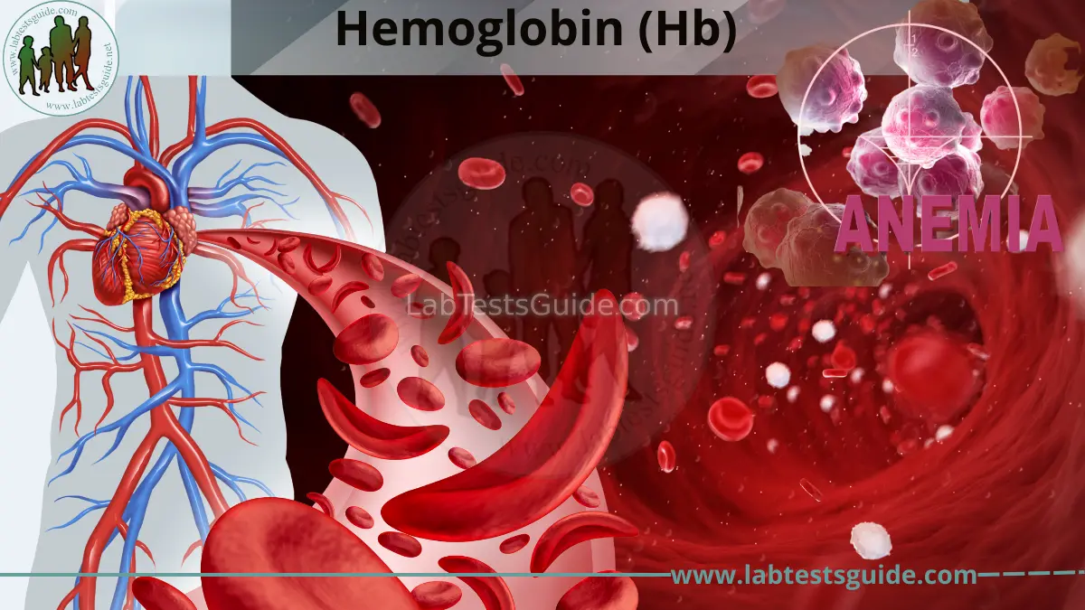
Hemoglobin (Hb) is an iron-containing respiratory protein found in erythrocytes (RBCs) responsible for the transport of oxygen from lungs to tissues and carbon dioxide back to the lungs. Clinically, hemoglobin measurement represents the concentration of Hb in whole blood and is a key indicator of oxygen-carrying capacity.
Clinical Importance
Hemoglobin estimation is essential for diagnosing and monitoring anemia, polycythemia, and blood loss conditions. It directly reflects the oxygen delivery capacity of blood and is routinely used in pre-surgical evaluation, pregnancy monitoring, and chronic disease management.
- Diagnosis of iron-deficiency and nutritional anemia
- Monitoring chronic kidney disease and chemotherapy
- Assessment of acute and chronic blood loss
Role in Modern Laboratory Medicine
Modern hematology analyzers determine hemoglobin using SLS-Hemoglobin method, cyanmethemoglobin (reference method), or non-cyanide spectrophotometric techniques. These methods ensure high accuracy, rapid reporting, and standardization across automated platforms.
Overview of the Test
Specimen: Whole blood (venous) collected in K₂EDTA tube.
Hemoglobin is measured as part of CBC or as a standalone test using automated
hematology analyzers to determine total Hb concentration in g/dL.
Basic Physiology & Scientific Background
Hemoglobin physiology reflects the integrated function of oxygen transport, iron metabolism, erythropoiesis, and tissue oxygen demand. Its concentration directly represents the oxygen-carrying capacity of blood and systemic oxygen delivery efficiency.
RBC–Hemoglobin Relationship
Hemoglobin is synthesized during erythropoiesis in bone marrow and packaged into mature erythrocytes. Each RBC contains approximately 270 million Hb molecules, making RBC count and Hb concentration physiologically interdependent. Any disruption in erythroid maturation directly alters Hb levels and oxygen delivery.
Iron Metabolism
Hemoglobin synthesis depends on iron availability, absorbed in the intestine and stored as ferritin. Hepcidin regulates iron absorption and release. Iron deficiency leads to reduced Hb synthesis, resulting in microcytic hypochromic anemia.
Oxygen Transport Physiology
Hemoglobin binds oxygen reversibly in the lungs and releases it in peripheral tissues based on partial pressure gradients (pO₂). The oxygen dissociation curve is influenced by pH, CO₂, temperature, and 2,3-BPG, regulating tissue oxygen delivery.
Erythropoietin Regulation
The kidney senses hypoxia and secretes erythropoietin (EPO), stimulating red cell and hemoglobin production in bone marrow. Chronic kidney disease reduces EPO output, leading to decreased Hb levels and normocytic anemia.
Hemoglobin Variants
Genetic mutations in globin chains produce abnormal hemoglobin such as HbS (sickle cell disease) and thalassemias. These disorders alter RBC structure, lifespan, and oxygen affinity, resulting in chronic hemolytic or ineffective erythropoiesis.
Tissue Oxygen Demand
Hemoglobin levels must match tissue oxygen demand. Conditions like fever, pregnancy, sepsis, and hyperthyroidism increase oxygen consumption, revealing latent anemia. Low Hb leads to tissue hypoxia, fatigue, and compensatory tachycardia.
Principle & Methodology
Hemoglobin estimation is based on spectrophotometric measurement of light absorbance after conversion of hemoglobin into a stable derivative. Accurate Hb analysis depends on complete lysis of RBCs and uniform color development proportional to Hb concentration.
Test Principle
Hemoglobin is measured by converting all forms of Hb into a stable colored compound (such as cyanmethemoglobin or SLS-Hb complex). The intensity of color formed is directly proportional to the Hb concentration and is measured using a spectrophotometer at a specific wavelength.
- RBC lysis: releases hemoglobin into solution
- Conversion: Hb → stable colored derivative
- Measurement: absorbance ∝ Hb concentration
Analytical Methodology
Hemoglobin estimation follows a standardized analytical workflow:
- Sample collection — EDTA whole blood
- Hemolysis — RBCs are lysed to release Hb
- Color development — conversion into measurable compound
- Photometric reading — absorbance measured at specific wavelength
Calibration: performed using standardized Hb calibrators traceable to reference methods.
Instrumentation
Hemoglobin measurement is performed using automated hematology analyzers or standalone photometers:
- Sysmex analyzers — SLS-Hb method
- Abbott Alinity — photometric Hb measurement
- Beckman Coulter DxH — cyanide-free Hb methods
Technology Used
Modern Hb estimation relies on integrated analytical technologies:
- Spectrophotometry — Hb absorbance measurement
- Cyanmethemoglobin method — reference standard
- SLS method — cyanide-free alternative
- Laboratory automation — integrated reporting systems
Specimen Requirements
Accurate hemoglobin estimation depends on proper pre-analytical handling of blood specimens. Even minor errors in collection or storage can significantly affect Hb concentration and lead to false low or falsely elevated results.
Specimen Type
Whole blood is required for hemoglobin estimation.
Preferred sample: venous blood collected from peripheral vein.
Capillary blood may be used for point-of-care Hb testing (fingerstick).
Collection Container
K₂EDTA or K₃EDTA (lavender-top) tube is required.
EDTA prevents coagulation and preserves hemoglobin integrity for accurate spectrophotometric analysis.
Avoid: heparin, citrate, or serum tubes (cause dilution errors).
Sample Volume
Adult: 2–3 mL whole blood is sufficient for Hb measurement.
Pediatric: 0.5–1 mL (microcollection acceptable).
Underfilling EDTA tube may cause incorrect anticoagulant ratio and affect results.
Transport Conditions
Transport at ambient temperature (18–25°C) within 2–4 hours.
If delay expected, store at 2–8°C and analyze as soon as possible.
Avoid: excessive shaking, heat exposure, and direct sunlight.
Storage Conditions
Room temperature: Hb stable up to 24 hours.
Refrigerated (2–8°C): stable up to 48 hours.
Freezing: not recommended due to RBC hemolysis risk.
Mix sample gently before analysis to avoid settling artifacts.
Analytical Stability
Hemoglobin is relatively stable compared to cellular parameters.
However, prolonged storage may cause slight concentration shifts due to RBC lysis or evaporation.
Best accuracy achieved when analyzed within 4–6 hours of collection.
Rejection Criteria
- Hemolyzed or visibly lysed sample
- Clotted blood in EDTA tube
- Wrong anticoagulant used
- Insufficient volume or underfilled tube
- Delayed analysis beyond acceptable time window
- Improper labeling or sample mismatch
Patient Preparation
Pre-analytical variables significantly affect hemoglobin accuracy. Proper preparation ensures reliable assessment of oxygen-carrying capacity and prevents false anemia or falsely elevated Hb values.
Fasting
Hemoglobin (Hb) testing does not require fasting.
- Food intake does not significantly alter Hb concentration in acute setting
- Hydration status may mildly influence plasma dilution
Normal diet and water intake are allowed before testing.
Medications
Document medications that may affect hemoglobin levels:
- Iron therapy — ↑ Hb over time
- Erythropoietin (EPO) — ↑ RBC & Hb
- Chemotherapy — ↓ Hb (bone marrow suppression)
- Anticoagulants/NSAIDs — may ↓ Hb via bleeding
Diet
Diet affects hemoglobin mainly in long-term erythropoiesis:
- Iron-rich diet — supports Hb synthesis
- Vitamin B12 & folate — prevents macrocytic anemia
- Malnutrition — chronic ↓ Hb
- Hydration status — dilutional changes in Hb
Exercise
Exercise causes temporary hemoconcentration affecting Hb:
- Hb may increase due to plasma volume loss (dehydration)
- Hydration restoration normalizes values
- Strenuous exercise may cause transient false elevation
Recommendation: Rest for 15–20 minutes before sampling.
Special Populations
- Pregnancy: physiological anemia due to plasma expansion (↓ Hb)
- Neonates: naturally higher Hb levels; capillary sampling preferred
- Elderly: higher anemia prevalence; chronic disease impact
- Chronic disease patients: CKD and inflammation lower Hb
- High altitude residents: physiologically elevated Hb levels
Hemoglobin (Hb) Test Procedure & Laboratory Workflow
Standardized laboratory workflow ensures accuracy, reliability, and traceability of Hemoglobin (Hb) results. Each step follows CLSI and ISO 15189 guidelines for optimal patient care.
Specimen Receipt
Preparation
Pre-analytical preparation before Hemoglobin analysis:
- Mixing — gentle inversion 8–10 times (EDTA blood)
- Hemolysis step — RBC lysis reagent added for Hb release
- Reagent preparation — SLS or Drabkin’s reagent (method dependent)
- QC material — 3 levels (low, normal, high Hb)
Analysis
Automated Hemoglobin measurement using spectrophotometry:
- Sample aspiration — 5–20 µL whole blood
- Hemoglobin conversion — Hb → HiCN (cyanmethemoglobin) or Hb-SLS complex
- Photometric reading — absorbance at specific wavelength (540–555 nm)
- Calculation — Hb concentration derived from calibration curve
Quality Checks
Quality assurance for Hb accuracy:
- Internal QC — daily Hb controls (low/normal/high)
- External QC — proficiency testing schemes
- Calibration verification — against reference standards
- Flagging system — hemolysis or lipemia interference alerts
Result Validation
Clinical validation of Hb results:
- Auto-validation — analyzer rule checks
- Manual review — abnormal Hb values confirmation
- Delta check — comparison with previous Hb results
- Critical Hb values — immediate notification
Reporting
Final Hemoglobin report generation:
- Electronic report — Hb value with reference range
- Interpretive comments — anemia / polycythemia correlation
- Critical Hb alert — immediate physician notification
- Archiving — long-term record storage
Hemoglobin (Hb%) Test Procedure
- Spectrophotometer / Colorimeter
- Calibrated Micropipettes (10–1000 µL)
- Disposable Tips & Cuvettes
- Test Tubes / Hematology Tubes (EDTA)
- Venous or Capillary Blood Sample
- Drabkin’s Reagent (Cyanmethemoglobin)
- Hemoglobin Standard (15.0 g/dL)
- Quality Control Materials (Low & High)
Drabkin’s Reagent (per Liter):
- Potassium ferricyanide: 200 mg
- Potassium cyanide: 50 mg
- KH₂PO₄ buffer: 140 mg
- Non-ionic detergent: 1 mL
Store at 15–25°C · Protect from light
| Wavelength | 540–546 nm |
| Cuvette Path | 1 cm |
| Temperature | 20–25°C |
| Measurement | Against Reagent Blank |
| Step | Blank | Std | Test |
|---|---|---|---|
| Drabkin’s Reagent | 5.0 mL | 5.0 mL | 5.0 mL |
| Standard (15.0) | — | 20 µL | — |
| Sample (Blood) | — | — | 20 µL |
| ⏱️ INCUBATION: Mix · 3–5 min · 20–25°C | |||
| Read Abs | Zero | Std Abs | Test Abs |
| Step | Blank | Calib | Test |
|---|---|---|---|
| Drabkin’s Reagent | 5.0 mL | 5.0 mL | 5.0 mL |
| Calibrator | — | 20 µL | — |
| Sample (Blood) | — | — | 20 µL |
| ⏱️ Hb = Test Abs × Factor | |||
| Result | Zero | Factor | Hb Conc. |
Linearity: 0–20 g/dL. Hemolyzed or lipemic samples may interfere with readings. Wavelength: 540–546 nm.
- Run low and high Hb controls daily
- Verify instrument calibration
- Maintain Levey-Jennings charts
- Hemolysis (↑ absorbance)
- Lipemia (↑ turbidity)
- Dehydration (false high Hb)
- Hematocrit (HCT) ratio
- RBC count & indices
- Iron studies (Ferritin)
- Oxygen transport capacity
- Anemia evaluation
- Polycythemia diagnosis
Hemoglobin (Hb) Normal Values
| Male | 14.0–17.5 g/dL 140–175 g/L |
| Female | 12.3–15.3 g/dL 123–153 g/L |
| Male | 12.3–16.6 g/dL 123–166 g/L |
| Female | 11.6–15.0 g/dL 116–150 g/L |
| Male | 13.0–17.0 g/dL 130–170 g/L |
| Female | 12.0–15.0 g/dL 120–150 g/L |
| Male | 14.0–18.0 g/dL 140–180 g/L |
| Female | 12.0–16.0 g/dL 120–160 g/L |
Hemoglobin (Hb) Unit Converter
• Hb% is calculated using the standard Haldane/Sahli scale where 100% = 14.6 g/dL.
• mmol/L is based on the Hemoglobin Tetramer molecular weight (~64,458).
Interpretation of Results
Hemoglobin (Hb) interpretation must be correlated with age, sex, hydration status, altitude, and clinical context. Abnormal values indicate disorders of red cell production, destruction, or loss.
High Hemoglobin
Above reference range — suggests increased red cell mass or hemoconcentration:
- Polycythemia vera: Primary myeloproliferative disorder
- Secondary polycythemia: Chronic hypoxia, COPD, high altitude, congenital heart disease
- Dehydration: Relative increase due to plasma volume loss
- Smoking: Increased carboxyhemoglobin leading to compensatory rise
Low Hemoglobin
Below reference range — indicates anemia or blood loss:
- Iron deficiency anemia: Most common cause
- Chronic disease anemia: Inflammation, infection, malignancy
- Acute blood loss: Trauma, GI bleeding, surgery
- Hemolytic anemia: RBC destruction (autoimmune, G6PD deficiency)
- Bone marrow suppression: Aplastic anemia, chemotherapy, leukemia
Normal Hemoglobin
Within reference range — suggests adequate oxygen-carrying capacity:
- Normal red cell production and turnover
- Adequate iron, B12, and folate status (usually)
- No significant ongoing blood loss or hemolysis
Note: Normal Hb does not exclude early anemia or hidden deficiency states — correlate with MCV, ferritin, and clinical symptoms.
Clinical Significance
Hemoglobin (Hb) reflects the oxygen-carrying capacity of blood and is a key marker for anemia, polycythemia, and systemic disease severity. Interpretation must be correlated with clinical findings and RBC indices.
Hemoglobin Abnormalities
- ↑ Hemoglobin: Polycythemia vera, secondary polycythemia (COPD, chronic hypoxia, high altitude), dehydration, smoking
- ↓ Hemoglobin: Anemia — iron deficiency, B12/folate deficiency, hemolysis, chronic disease, bone marrow failure
- ↓ Hb with ↓ MCV: Microcytic anemia — iron deficiency, thalassemia
- ↓ Hb with ↑ MCV: Macrocytic anemia — B12 deficiency, folate deficiency, alcoholism
- Iron deficiency anemia: ↓ Hb, ↓ MCV, ↑ RDW, low ferritin
- Thalassemia: ↓ Hb, very ↓ MCV, normal/high RBC count
- Chronic kidney disease: ↓ Hb due to ↓ erythropoietin
- Acute blood loss: Trauma, GI bleeding, surgery
- Low Hb: Associated with increased mortality in CKD, heart failure, and chronic respiratory disease
- Severe anemia: Reduced tissue oxygenation and organ dysfunction risk
- High Hb: Increased thrombotic risk in polycythemia states
Associated Diseases & Conditions (Hemoglobin Test)
The Hemoglobin (Hb) test is closely associated with disorders affecting red blood cells, oxygen-carrying capacity, and systemic oxygen delivery. It is essential for diagnosing anemia, polycythemia, and hemoglobinopathies.
Red Cell & Hb Disorders
- Iron deficiency anemia — most common cause of low Hb, microcytosis, ↓ ferritin
- Vitamin B12 deficiency — macrocytic anemia, neurological symptoms
- Folate deficiency — megaloblastic anemia, poor diet or malabsorption
- Hemolytic anemia — ↑ LDH, ↓ haptoglobin, jaundice
- Aplastic anemia — bone marrow failure, pancytopenia
- Anemia of chronic disease — inflammation-related Hb reduction
- Polycythemia vera — JAK2 mutation, ↑ Hb/Hct
- Secondary erythrocytosis — COPD, sleep apnea, high altitude, cyanotic heart disease
- Sickle cell disease — HbS, vaso-occlusive crises
- Thalassemia — α/β chain defects, microcytic anemia
Infectious & Hematologic Conditions
- Malaria — hemolysis causing severe anemia and ↓ Hb
- Bacterial infections — chronic disease anemia, inflammation
- Viral infections — bone marrow suppression, transient ↓ Hb
- Sepsis — hemolysis + anemia of critical illness
Bleeding & Platelet Disorders
- Gastrointestinal bleeding — major cause of acute ↓ Hb
- Trauma / hemorrhage — acute blood loss anemia
- Menorrhagia — chronic iron deficiency anemia
- Immune thrombocytopenia (ITP) — bleeding risk, ↓ Hb in severe cases
- DIC — consumption coagulopathy, hemorrhage
- Thrombocytopenia — mucosal bleeding, petechiae
Systemic & Organ Disorders
- Chronic kidney disease — ↓ erythropoietin → ↓ Hb
- Cirrhosis — hypersplenism, anemia, bleeding tendency
- Alcoholic liver disease — macrocytosis + ↓ Hb
- Diabetes mellitus — chronic disease anemia, CKD-related Hb drop
- Malnutrition — iron, B12, folate deficiency anemia
Quality Control & Laboratory Considerations (Hemoglobin Test)
Hemoglobin (Hb) testing requires strict analytical control to ensure accuracy, especially in diagnosing anemia, polycythemia, and monitoring therapy response. Proper quality systems reduce analytical variation and improve diagnostic reliability.
Internal Quality Control (IQC) for Hb
Continuous monitoring of Hb analyzer performance using control materials to ensure analytical precision and accuracy.
- 📊 Low Hb control: Anemic range verification
- 📊 Normal Hb control: Reference range validation
- 📊 High Hb control: Polycythemic range check
- 📊 Matrix: Whole blood-based stabilized controls
- 📊 Methods: Cyanmethemoglobin, SLS-Hb, HPLC, POCT
- 📊 Levey-Jennings charts for trend analysis
- 📊 Westgard rules for rejection criteria
- 📊 CV target: <2–3% for automated analyzers
External Quality Assessment (EQA)
Inter-laboratory comparison programs ensure Hb results are standardized and traceable.
- 🌐 CAP PT: Hemoglobin proficiency testing
- 🌐 UK NEQAS: Hematology external quality scheme
- 🌐 WHO standards: Reference Hb calibration alignment
- 📋 Comparison against peer group mean
- 📋 Acceptable limit: ±2 SD or ±5% variation
- 🚫 Outlier detection triggers corrective action
Hemoglobin Calibration
Calibration ensures accurate conversion of optical or electrical signals into true hemoglobin concentration.
- ⚙️ Reference method: Cyanmethemoglobin standard (gold standard)
- ⚙️ Modern analyzers: SLS-Hb photometric calibration
- ⚙️ POCT devices: Factory-calibrated cuvette systems
- ⚙️ Multi-point calibration: Low–normal–high range mapping
- ✅ Calibrator traceability to IFCC/ICSH standards
- 🔄 Recalibration after reagent lot change
- 📊 Drift monitoring using control charts
Method Validation for Hb
Ensures Hb assay performance meets clinical requirements for diagnosis and monitoring.
- 📊 Precision: Repeatability & reproducibility (CV%)
- 📊 Accuracy: Comparison with reference cyanmetHb method
- 📊 Linearity: 3–20 g/dL (typical analytical range)
- 📊 Interference: Lipemia, hemolysis, bilirubin effects
- 📋 CLSI EP05 / EP06 guidelines
- 📋 ≥40 patient sample comparison studies
- 📋 Reference interval verification (healthy population)
Hb Analyzer Maintenance
Regular maintenance ensures optical stability, reagent integrity, and accurate Hb measurement.
- 🔹 Blank check (distilled water/zeroing)
- 🔹 Clean cuvettes or flow system
- 🔹 Run QC controls (low/normal/high)
- 🔹 Optical path cleaning (photometer alignment)
- 🔹 Reagent stability check (Drabkin/SLS reagents)
- 🔹 Carryover and drift assessment
- 🔹 Full calibration verification
- 🔹 Preventive service by manufacturer
- 🔹 Performance qualification (PQ)
Sources of Error & Limitations (Hemoglobin Test)
Understanding sources of error and limitations in hemoglobin (Hb) testing is essential for accurate interpretation of anemia and erythrocyte disorders. Errors may occur at any stage of the total testing process.
Pre-analytical Errors
Most frequent source of Hb variation, often due to sample collection, patient condition, or handling.
- Dehydration — falsely ↑ Hb (hemoconcentration)
- Overhydration — falsely ↓ Hb (hemodilution)
- Smoking — ↑ carboxyhemoglobin, pseudo-elevated Hb
- High altitude — physiological polycythemia
- Recent transfusion — mixed population of RBCs
- Hemolysis — in vitro RBC destruction alters results
- Incorrect anticoagulant — EDTA required for CBC/Hb
- Clotted sample — underestimation of Hb
- Inadequate mixing — uneven cell distribution
- Delayed analysis — RBC swelling/lysis
- Temperature extremes — cell membrane instability
- Improper storage — changes in hematologic indices
Analytical Errors
Errors occurring during instrument measurement of hemoglobin.
- Photometric interference — turbidity affects absorbance
- Optical misalignment — inaccurate Hb reading
- Calibration drift — slope/intercept deviation
- Clogged flow cell — inaccurate sample aspiration
- Lipemia — falsely ↑ or ↓ Hb due to turbidity
- Hyperbilirubinemia — spectral interference
- Carboxyhemoglobin — seen in CO poisoning
- Hemoglobin variants — HbS, HbC affect measurement accuracy
- Reagent instability — expired or degraded reagents
- Incomplete RBC lysis — underestimation of Hb
- Carryover effects — contamination from prior sample
Post-analytical Errors
Errors occurring after Hb result generation.
- Incorrect result entry — transcription errors in Hb value
- LIS mismatch — wrong patient assignment
- Delayed reporting — impacts anemia management
- Ignoring clinical context — Hb alone not diagnostic
- Failure to correlate with RBC indices
- Overreliance on single Hb value
Test Limitations
Inherent limitations of Hb testing in diagnosis and monitoring.
- Not disease-specific — cannot diagnose anemia type alone
- Physiological variation — age, sex, altitude differences
- Pregnancy dilutional anemia — physiological ↓ Hb
- Acute blood loss delay — Hb may remain initially normal
- Limited detection of variants — HbS/HbC interference
- Linearity constraints — extreme values require dilution
- Instrument dependency — method variability across analyzers
- Requires CBC correlation — RBC indices essential
- Cannot assess iron status alone
- Trend analysis required — single value insufficient
Clinical Applications
The Hemoglobin (Hb) test is a fundamental component of hematology used across nearly all clinical settings. It plays a central role in diagnosing oxygen-carrying disorders, especially anemia and polycythemia, and guiding patient management.
Hospital Use
Routine hospital testing for diagnosis, monitoring, and discharge assessment.
- Admission workup — baseline Hb in all patients
- Post-operative monitoring — detect hidden bleeding
- Discharge evaluation — ensure hematologic stability
- Anemia — diagnosis and classification
- Thalassemia — severity assessment
- Sickle Cell Disease — disease monitoring
- Cardiology — ischemia risk assessment
Screening Programs
Early detection of hematologic and nutritional disorders in populations.
- Routine health check-ups — anemia screening
- Community screening — iron deficiency, hemoglobinopathies
- Occupational screening — chronic exposure monitoring
- Pregnancy — anemia screening in antenatal care
- Elderly — chronic disease anemia detection
- Newborns — congenital hemoglobin disorders
Emergency Medicine
Rapid Hb assessment is critical in acute and life-threatening conditions.
- Acute blood loss — trauma, GI hemorrhage
- Severe anemia — symptomatic hypoxia
- Hemolytic crisis — transfusion reactions
- Shock states — oxygen delivery impairment
- Transfusion decision — Hb threshold-based care
- Risk stratification — severity assessment
- Admission priority — ICU vs ward allocation
ICU Use
Continuous monitoring of oxygen-carrying capacity in critically ill patients.
- Serial Hb testing — trend monitoring
- Blood loss tracking — surgical/trauma ICU
- Transfusion guidance — ICU thresholds
- Sepsis — anemia of inflammation
- Multi-organ failure — hematologic decline
- Post-surgery — occult bleeding
- Burn patients — hemolysis and fluid shifts
Monitoring Therapy
Hemoglobin response monitoring guides treatment effectiveness and dose adjustment.
- Iron deficiency — Hb recovery tracking
- Ferritin correlation — storage evaluation
- Oral/IV iron — response monitoring
- Chronic kidney disease — Hb target maintenance
- EPO response — reticulocyte rise
- Dose adjustment — avoid overcorrection
- Pre/post Hb — effectiveness check
- Massive transfusion — resuscitation monitoring
- Chronic transfusion — iron overload risk
Critical Values & Alert Conditions (Hemoglobin)
Hemoglobin (Hb) critical values indicate life-threatening anemia or polycythemia requiring urgent clinical intervention such as blood transfusion or immediate evaluation.
Severe Anemia (Low Hemoglobin)
- Adults (general): ≤ 7.0 g/dL — Consider urgent transfusion
- Life-threatening: ≤ 5.0 g/dL — Immediate transfusion required
- Pediatric (severe): ≤ 6–7 g/dL — Urgent evaluation
- 🩸 Acute or chronic blood loss
- 🧬 Iron deficiency anemia
- 🧪 Hemolytic anemia
- 🦴 Bone marrow suppression
Polycythemia / Elevated Hemoglobin
- Adult male: ≥ 20.0 g/dL — Hyperviscosity risk
- Adult female: ≥ 18.5 g/dL — Evaluate for polycythemia
- 🫁 Chronic hypoxia (COPD, high altitude)
- 🧬 Polycythemia vera
- 💊 Erythropoietin excess
- 🚬 Smoking-related secondary polycythemia
Urgent Clinical Conditions
- Acute bleeding: rapid Hb drop (>2 g/dL) — Suspect hemorrhage
- Cardiovascular instability: Hb ≤ 7 g/dL with symptoms — Tachycardia, hypotension
- Neurologic symptoms: dizziness, syncope — Tissue hypoxia
- ❤️ Cardiac disease patients (Hb < 8 g/dL critical risk)
- 🫁 COPD / chronic lung disease
- 🤰 Pregnancy (physiologic anemia risk)
Laboratory Alert & Notification
- 🔬 Repeat analysis for critical Hb values
- 🧪 Check sample integrity (hemolysis, clotting)
- 📊 Correlate with CBC parameters
- 📞 Immediate phone call to clinician
- 🧾 Document time of communication
- 📤 Record in LIS system
- ⚠️ Escalate if no response received
Special Considerations (Hemoglobin Test)
Hemoglobin interpretation varies significantly across age, sex, pregnancy status, altitude, and clinical conditions. Accurate diagnosis requires context-aware evaluation rather than fixed reference ranges alone.
Pregnancy
Physiological hemodilution causes a natural decline in hemoglobin levels during pregnancy.
- Hemoglobin: ↓ ~1–2 g/dL — dilutional (plasma volume expansion)
- Iron demand: ↑ significantly — increased maternal & fetal needs
- Lower limit (WHO): < 11 g/dL — anemia in pregnancy
- Iron deficiency anemia: most common cause
- Supplementation: routine iron + folate recommended
- Monitoring: antenatal Hb screening at booking & 28 weeks
Pediatrics
Age-dependent hemoglobin variation is significant from neonates to adolescents.
- Neonates: 14–24 g/dL — physiologically high at birth
- Infants (2–3 months): physiologic nadir — “physiologic anemia of infancy”
- Children: 11–15 g/dL — gradual normalization
- Iron deficiency is the most common cause of anemia
- Minimize blood draw volume to prevent iatrogenic anemia
- Screen for hemoglobinopathies in high-risk populations
Elderly Patients
Mild hemoglobin decline is common due to chronic disease and aging physiology.
- Hemoglobin: mildly decreased — often 11–13 g/dL
- Causes: chronic kidney disease, inflammation, malnutrition
- ESRD risk: reduced erythropoietin production
- Evaluate for iron, B12, folate deficiency
- Screen for occult malignancy if unexplained anemia
- Consider CKD-related anemia management
Altitude & Lifestyle
Environmental and lifestyle factors significantly influence hemoglobin levels.
- High altitude: ↑ Hb — compensatory erythrocytosis
- Smoking: ↑ Hb — chronic hypoxia response
- Athletes: plasma volume expansion → pseudoanemia
- Adjust reference ranges for altitude residents
- Differentiate true anemia vs dilutional pseudoanemia
- Consider oxygen saturation context
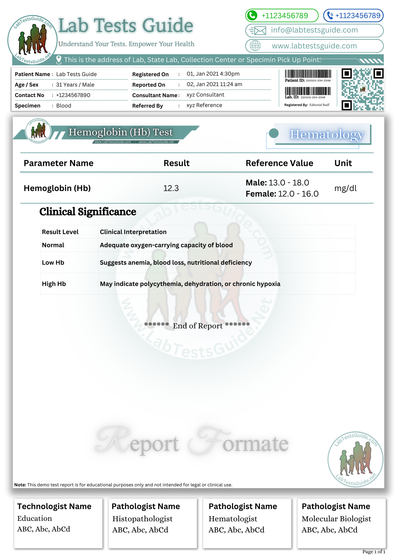
Download CBC Lab Report Format
Get the demo report format for Complete Blood Count (CBC) in your preferred format. These templates are fully editable, professionally designed, and ready to use.
Frequently Asked Questions (Hb Test)
Common questions about the Hemoglobin (Hb) test from patients and learners. Click on any question to view the answer.
The hemoglobin (Hb) test measures the amount of hemoglobin protein in red blood cells, which is responsible for oxygen transport in the blood.
No fasting is required for a hemoglobin test. It can be performed at any time of the day.
Normal ranges are approximately 13–18 g/dL in men and 12–16 g/dL in women, but values may vary slightly by laboratory.
Low Hb is commonly caused by iron deficiency anemia, blood loss, vitamin deficiencies (B12/folate), chronic disease, or bone marrow disorders.
High Hb may occur due to dehydration, smoking, high altitude living, chronic hypoxia, or conditions like polycythemia vera.
Yes, hemoglobin is a key parameter of the Complete Blood Count (CBC) and is used to assess anemia and overall blood health.
The cyanmethemoglobin (Drabkin) method is considered the reference method for hemoglobin estimation in laboratory practice.
Yes, dehydration can falsely increase hemoglobin levels due to reduced plasma volume (hemoconcentration).
The Hb test is usually completed within 1–2 hours in routine laboratory settings.
Hemoglobin is essential because it reflects the blood’s oxygen-carrying capacity and helps detect anemia, bleeding disorders, and other hematological conditions.
Common methods include SLS-Hemoglobin (Sodium Lauryl Sulfate), cyanmethemoglobin (reference method), and spectrophotometric measurement in automated hematology analyzers.
Factors include improper anticoagulant ratio, hemolysis, delayed analysis, sample dilution, and poor mixing of EDTA tubes.
Hemolysis releases intracellular hemoglobin into plasma, which may cause falsely elevated Hb results depending on the analytical method used.
Hemoglobin variants (HbS, HbC, HbE) may affect accuracy in some analyzers and may require HPLC or electrophoresis for confirmation.
Quality control includes daily internal QC with control materials, calibration checks, and participation in external quality assurance programs (EQA).
Critical Hb values (<5 g/dL or >20 g/dL) require immediate clinical notification due to risk of severe anemia or polycythemia complications.
Modern analyzers use specific chemical conversion methods (e.g., SLS method) and wavelength-specific spectrophotometry to minimize interference.
Hemoglobin is used to monitor anemia in chronic kidney disease, cancer therapy, inflammatory diseases, and response to treatment.
References:
- WHO Anemia Detection Guide – WHO – (Accessed on Mar 09, 2026)
- Clinical Hematology Principles and Procedures – Archive – (Accessed on Mar 09, 2026)
- Tulip Diagnostics Hemoglobin Reagent Insert – Tulip Diagnostic – (Accessed on Mar 09, 2026)

6 Comments