Dental technology has undergone a profound transformation over the past two decades, and nowhere is this more evident than in the area of diagnostic imaging. Digital X-rays have largely replaced traditional film-based radiography in modern dental practices, offering faster results, lower radiation exposure, and significantly enhanced diagnostic capabilities. Whether you are attending a routine check-up or presenting with a specific concern, digital X-rays are likely to play a central role in your dental examination – and understanding how they work can help you appreciate the quality of care you are receiving.
What Are Digital X-Rays?
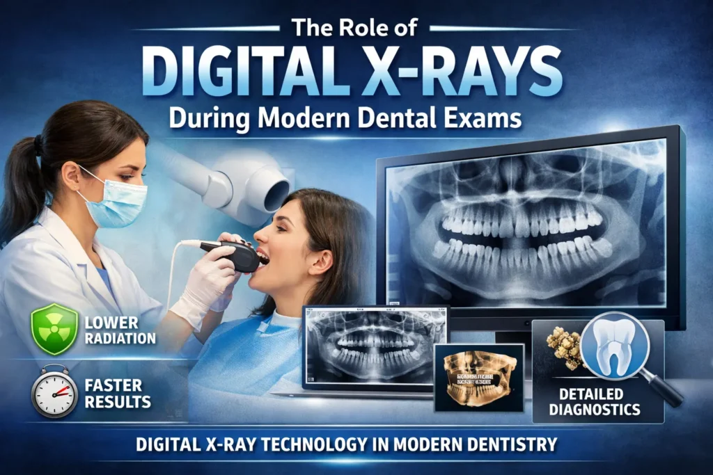
Digital X-rays, also known as digital radiographs, use electronic sensors rather than traditional photographic film to capture images of the teeth, gums, jawbone, and surrounding structures. When X-ray beams pass through oral tissues, the sensor records varying levels of absorption and transmits the data directly to a computer, where images are rendered in seconds.
There are two primary types of digital dental X-rays. Intraoral X-rays are taken with the sensor placed inside the mouth, producing detailed images of individual teeth and the supporting bone. Extraoral X-rays, such as panoramic and cephalometric images, capture broader views of the entire jaw and skull, making them especially useful for orthodontic assessment and oral surgery planning.
Digital vs. Traditional Film X-Rays
While both technologies rely on radiation to produce images, digital X-rays offer several meaningful advantages over conventional film-based methods:
Reduced Radiation Exposure
One of the most frequently cited benefits of digital radiography is the significant reduction in radiation dose. Digital X-rays typically emit up to 80% less radiation than traditional film X-rays, making them considerably safer for patients of all ages, including children and pregnant women requiring diagnostic imaging.
Faster Image Acquisition
Unlike film X-rays, which require chemical development that can take several minutes, digital images appear on the dentist’s screen within seconds. This speed allows the dentist to review findings with the patient in real time and make quicker clinical decisions.
Superior Image Quality and Manipulation
Digital images can be enhanced, magnified, and adjusted for contrast and brightness without loss of quality. This ability to manipulate images in real time improves diagnostic accuracy, allowing dentists to identify subtle issues – such as early-stage cavities or hairline fractures – that might be missed on traditional film.
Environmentally Friendly
Traditional film processing requires chemical developers and fixers that pose disposal challenges. Digital X-rays eliminate the need for these chemicals entirely, making the process cleaner and more environmentally sustainable.
What Can Digital X-Rays Detect?
Digital X-rays give dentists a comprehensive view of oral structures that are simply invisible to the naked eye. During a routine dental exam, they are used to detect and monitor a wide range of conditions, including:
- Cavities (dental caries) – especially those developing between teeth or beneath existing fillings
- Bone loss associated with periodontal (gum) disease
- Infections, cysts, or abscesses at the root of a tooth
- Impacted teeth, including wisdom teeth that have not fully erupted
- Developmental abnormalities in children, such as missing or extra teeth
- Tumours or other pathological changes in the jaw and surrounding bone
- Root fractures or damage not visible during a clinical examination
- Changes in bone density that may indicate systemic conditions such as osteoporosis
The ability to detect these issues at an early stage is one of the most significant clinical advantages of digital radiography. Early diagnosis invariably leads to simpler, less invasive, and more cost-effective treatment.
How Often Should Dental X-Rays Be Taken?
The frequency of dental X-rays depends on the individual patient’s oral health history, age, and risk factors. For adults with no ongoing dental concerns and a history of good oral health, bitewing X-rays are typically recommended every 18 to 24 months. Patients with a higher risk of decay, gum disease, or other conditions may require imaging more frequently – sometimes every six to twelve months.
Children generally require X-rays more often than adults because their teeth and jaws are still developing and they are more susceptible to tooth decay. A full-mouth series of X-rays may be recommended for new patients to establish a comprehensive baseline, and panoramic images are commonly taken during adolescence to assess the development of wisdom teeth.
Safety Considerations
A common concern among patients is the safety of dental X-rays, particularly regarding radiation exposure. It is reassuring to note that the radiation dose from a full set of digital dental X-rays is comparable to the natural background radiation a person receives during a short aeroplane flight. Nonetheless, dental practices adhere to the ALARA principle – As Low As Reasonably Achievable – ensuring that X-rays are only taken when clinically justified.
Protective measures such as lead aprons and thyroid collars are routinely used to shield sensitive areas of the body. Patients should always inform their dentist if they are pregnant, as precautionary measures may be taken to defer non-urgent X-rays.
Reference: American Dental Association Council on Scientific Affairs (2006). “The Use of Dental Radiographs: Update and Recommendations.” Journal of the American Dental Association, 137(9), 1304–1312. This landmark ADA report establishes evidence-based guidelines for the appropriate use and frequency of dental radiographs, with specific recommendations tailored to patient age, risk profile, and clinical findings.
The Role of Digital X-Rays in Preventive Dentistry
Beyond diagnosis, digital X-rays serve a vital function in preventive care. By establishing a visual record of a patient’s oral structures over time, dentists can track subtle changes and intervene before conditions worsen. A small area of demineralisation detected on an X-ray may be monitored and managed with fluoride treatments rather than immediately requiring a filling – a clear example of how imaging supports minimally invasive dentistry.
Digital imaging also facilitates better communication between dental specialists. When a general dentist refers a patient to an orthodontist, periodontist, or oral surgeon, high-resolution digital images can be shared electronically in seconds, eliminating the need for duplicate X-rays and ensuring all providers have access to the same diagnostic information.
Cone Beam CT: The Next Step in Digital Imaging
For complex cases, many modern dental practices have adopted Cone Beam Computed Tomography (CBCT), a three-dimensional imaging system that provides detailed cross-sectional views of the teeth, bone, soft tissue, nerve pathways, and sinuses in a single scan. CBCT is particularly valuable in implant planning, endodontic treatment, and the management of jaw disorders, offering a level of diagnostic detail that two-dimensional X-rays cannot match.
While CBCT delivers a higher radiation dose than standard digital X-rays, it remains far below the threshold associated with adverse health effects and is used selectively when the clinical benefit justifies its use.
What to Expect During a Digital X-Ray
The process of taking digital X-rays is quick, straightforward, and painless. A dental hygienist or assistant will position a small sensor inside your mouth, ask you to bite down gently, and take the exposure – which lasts only a fraction of a second. The image appears on the chairside monitor almost immediately, and your dentist can review findings with you during the same appointment. A full set of bitewing X-rays typically takes no more than ten to fifteen minutes.
Conclusion
Digital X-rays are no longer a luxury reserved for specialist practices – they are a standard component of comprehensive dental care. Their ability to detect hidden problems early, reduce radiation exposure, and support a more collaborative and preventive approach to dentistry makes them an indispensable tool in the modern dental exam. For patients and practitioners alike, embracing digital radiography means embracing a higher standard of oral health. To learn more about how digital imaging and preventive dental care can benefit you, visit dental-matters.com. Staying informed is the first step toward a healthier smile.