🫀 Arteries vs. Veins
A Complete Vascular Atlas
Arteries: High Pressure
Thick, muscular, elastic walls withstand 80–120 mmHg. Carry oxygenated blood (except pulmonary). Pulse propagation.
Veins: Low Pressure
Thin walls, larger lumen, valves prevent backflow. Capacitance vessels store ~70% blood volume. 2–10 mmHg.
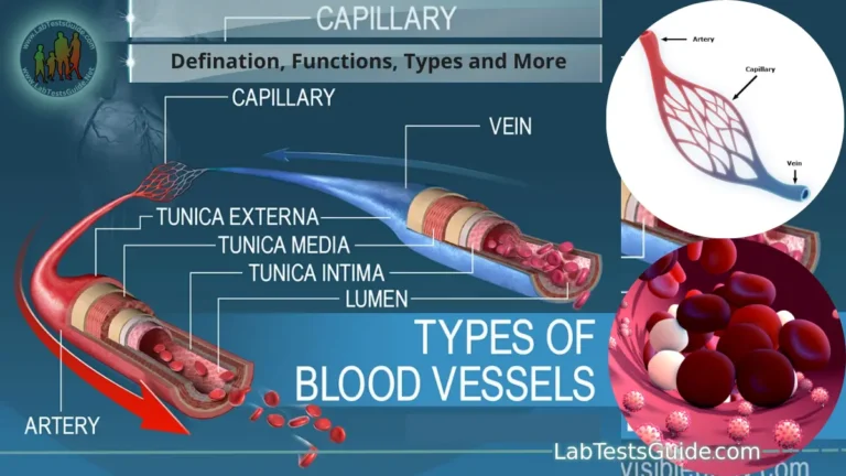
Histology Snapshot
Arteries: abundant tunica media (smooth muscle & elastin). Veins: thinner media, prominent tunica adventitia.
Clinical Relevance
Atherosclerosis, aneurysm (arteries) vs DVT, varicose veins, chronic venous insufficiency.

ARTERIES ⚡
- Direction: Away from heart → tissues
- Wall thickness: 1–4 mm (thick media)
- Lumen shape: Round, smaller diameter
- Elastic fibers: Abundant (elastic arteries)
- Smooth muscle: Highly developed (vasoconstriction)
- Valves: Absent (except semilunar valves at heart)
- Blood color: Bright red (oxygenated)
- Pulse: Palpable, rhythmic expansion
- Pressure: High pulsatile flow (80-120 mmHg)
VEINS 🌊
- Direction: Toward the heart (periphery → center)
- Wall thickness: ~0.5 mm (thin, collapsible)
- Lumen shape: Irregular, larger diameter
- Elastic fibers: Few, more collagen
- Smooth muscle: Sparse, but responsive to sympathetic tone
- Valves: Present (especially limbs) — bicuspid leaflets
- Blood color: Dark red (deoxygenated)
- Pulse: Non-palpable, venous hum possible
- Pressure: Low (2-10 mmHg), influenced by skeletal muscle pump

Comparative Hemodynamic Metrics
📐 Hemodynamics & Vascular Resistance
Flow is governed by Ohm’s Law of Hydrodynamics (ΔP = Q × R) and the Hagen-Poiseuille Equation. Because resistance is inversely proportional to the radius to the fourth power, small changes in arterial diameter via vasoconstriction massively shift blood pressure.

🔹 Arterial System Compliance
Elastic arteries (aorta, carotids) dampen pressure oscillations — Windkessel effect. Compliance = ΔVolume/ΔPressure. Arterial stiffness increases with age and atherosclerosis.
🔻 Venous Return Mechanics
Venous pressure gradient (≈7–10 mmHg) + skeletal muscle pump + respiratory thoracic pump. Valves crucial against gravity. Venous pooling leads to edema.
⚙️ Poiseuille’s Law & Radius
Resistance ∝ 1/r⁴. Arterioles are major resistance vessels. Veins offer low resistance but high capacitance — sympathetic venoconstriction shifts blood centrally.
🧬 Microanatomy: Tunics Compared

📌 Tunica Intima (Inner)
Artery: Endothelium + internal elastic lamina (prominent wavy membrane). Prevents clotting & regulates tone.
Vein: Thin endothelium, occasional internal elastic lamina absent or fragmented. Forms valve leaflets.
💪 Tunica Media (Middle)
Artery: 40–70 layers of smooth muscle in muscular arteries; elastic lamellae in elastic arteries. Rich in vasa vasorum.
Vein: 10–20 sparse smooth muscle layers, less elastin, more collagenous support.
🔗 Tunica Adventitia (Outer)
Artery: Relatively thin, collagen & elastic fibers. Contains nervi vasorum.
Vein: Thickest layer — longitudinal collagen bundles, supports large capacitance and prevents overdistension.
🩸 Oxygenation Misconceptions: Pulmonary Circuit
Pulmonary arteries carry deoxygenated blood (from right ventricle to lungs), while pulmonary veins return oxygenated blood to left atrium. This is the classic exception reinforcing functional classification: arteries always conduct blood away from the heart; veins conduct toward the heart regardless of oxygen content. The umbilical circulation in fetus presents another exception.

Pulmonary Circuit
Pulmonary Artery: Carries deoxygenated blood from RV to Lungs.
Pulmonary Veins: Return oxygenated blood to the LA.
Umbilical Circuit
Umbilical Arteries: Channel waste-laden blood to the placenta.
Umbilical Vein: Delivers oxygenated, nutrient-rich blood to the fetus.
🦵 Venous Valves & Skeletal Muscle Pump: Clinical Implications
Deep veins of lower extremities contain bicuspid valves preventing retrograde flow. During calf contraction, venous pressure rises to ~20–30 mmHg, propelling blood upward. Incompetent valves → varicose veins, venous reflux disease, chronic edema. Compared to arterial system, valves are absent except in heart (aortic/pulmonary). The soleus muscle is particularly important as the “peripheral heart.”

- Unidirectional Valves: Semilunar folds of intima that open toward the heart and snap shut to prevent retrograde pooling.
- Skeletal Muscle Pump: Contraction of deep leg muscles (e.g., Calf/Soleus) squeezes veins, propelling blood upward.
- Respiratory Thoracic Pump: Inhalation creates negative pressure in the chest, pulling venous blood up like a vacuum.
🔎 Arterial Disorders
Atherosclerosis — plaque formation in intima → stenosis, ischemia. Aneurysm — focal dilation >50% normal diameter, risk of rupture. Arteritis (Takayasu, giant cell). Peripheral Artery Disease (PAD) causing claudication.
🩹 Venous Disorders
Deep Vein Thrombosis (DVT): Virchow’s triad (stasis, hypercoagulability, endothelial injury). Varicose veins: superficial venous insufficiency. Chronic venous insufficiency: skin changes, lipodermatosclerosis, venous ulcers.
🔬 Diagnostic Tools
Ankle-Brachial Index (ABI) for PAD; Duplex ultrasound (arterial/venous flow). Venography gold standard for DVT. Arteriography for stenosis. CT Angiography for aneurysms.
Differential Diagnosis & Pathophysiology

Arterial Conditions
Atherosclerosis: Plaque buildup in the intima causing stenosis. A disease unique to arteries due to high pressure and shear stress.
Aneurysm: Focal dilation caused by weakened wall elastic layers. Risk of rupture.
PAD/Ischemia: Claudication (pain on walking), decreased pulses, pale/cyanotic skin, and pain relieved by hanging legs down.
Venous Conditions
DVT: Clot formation in deep veins. Risks: Virchow’s triad (stasis, endothelial injury, hypercoagulability). Risk of Pulmonary Embolism.
Varicosities: Valve failure leads to blood pooling, stretching superficial walls (tortuous, enlarged veins).
Chronic Insufficiency: Edema, brawny discoloration (hemosiderin staining), and relief with elevation.
Professional Deep-Dives: Academic Breakdown
Baroreceptor Matrix
Nervous system monitoring via stretch receptors in high-pressure arterial zones (Carotid sinus, Aortic arch). Veins lack these, relying on volume receptors in the Right Atrium.
Forensic Profile
Post-mortem: Arteries are usually empty and firm (elastic recoil). Veins remain filled with dark, stagnant blood and collapse.
Vasa Vasorum
Large vessels are too thick for diffusion. They have their own nutrient supply (vessels of vessels), more prominent in large veins due to lower luminal O₂.
📊 Diagnostic Benchmarks & Numerical Values
9. Clinical Interventions & Traumatology
Phlebotomy & IV Access: Primarily uses superficial veins because they are easy to visualize, have lower pressure (limiting bleeding after withdrawal), and have thinner walls.
Emergency Bleeding: Arterial bleeding is bright red and “spurts” in sync with heartbeats (requires tourniquet). Venous bleeding is dark maroon and flows in a steady, slow stream.
Surgical Grafting: The Great Saphenous Vein is often harvested for CABG. When moved to the heart, it undergoes “arterialization,” thickening its walls to handle the new high-pressure environment.
🧠 Embryology & Comparative Anatomy
From a developmental perspective, arteries and veins derive from the mesodermal layer. During angiogenesis, primitive vascular networks differentiate: arteries acquire smooth muscle from neural crest (in arch vessels) or mesoderm, while venous identity is regulated by COUP-TFII transcription factor. Arterial specification involves Notch signaling and ephrin-B2, whereas venous endothelium expresses Eph-B4. This molecular distinction explains why arteries and veins cannot interconvert under normal conditions. Understanding this aids in vascular malformation treatments.
⚡ Quick Self-Assessment (students & practitioners)
❓ Q1
Why do veins have a larger lumen compared to companion arteries?
✅ Lower resistance to accommodate large volume at low pressure.
❓ Q2
What prevents venous blood from pooling in the lower limbs when standing?
✅ Valves, skeletal muscle pump, and sympathetic tone.
❓ Q3
Which layer is significantly thicker in arteries vs veins?
✅ Tunica media (smooth muscle & elastin).
❓ Q4
Name the vessel that carries deoxygenated blood away from the heart.
✅ Pulmonary artery.
❓ Q5
Which vessel is the primary site of vascular resistance?
✅ Arterioles (Smallest arterial branches)./p>
❓ Q6
How does aging affect arterial vs venous structures?
✅ Arteries: Loss of elastin (stiffening). Veins: Loss of valve elasticity (edema)./p>
❓ Q7
Why do veins have a larger lumen than arteries?
✅ To accommodate large volumes at low resistance./p>
🏥 Bedside Correlations: When Arteries & Veins Collide
Arteriovenous fistula (AVF): Abnormal connection between artery and vein, bypassing capillaries. Can be congenital or iatrogenic (dialysis access). Leads to high-output cardiac failure, local venous distension, and audible bruit. Surgical repair or endovascular management required. Conversely, varicose veins: incompetent superficial veins, treatment: endovenous laser or sclerotherapy.
📈 Modern Imaging & Hemodynamic Parameters
Doppler waveforms: Arterial — triphasic (high resistance vessels) vs biphasic/monophasic (stenosis). Venous — phasic with respiration, augmentation with distal compression. Intima-media thickness (IMT) >0.9 mm carotid artery suggests subclinical atherosclerosis. Venous refill time >20s rules out significant valvular incompetence. Point-of-care ultrasound (POCUS) is now standard for DVT evaluation.
🧾 Summary Mnemonic (Easy Recall)
“A-V A-V” → Arteries: Away, thick wall, high pressure, no valves (except heart). Veins: Valves, low pressure, large lumen, Volume reservoir.
Histology hack: Arteries = muscular/elastic; Veins = thin walled with valves in limbs. Oxygenation ≠ Naming.
Additional: “Red Arteries Rush Away, Blue Veins Return Back”
📘 High-Yield Board Review (USMLE / COMLEX / NCLEX)
➜ Which vessel has the highest proportion of smooth muscle? Muscular arteries (e.g., femoral, radial).
➜ What effect does positive pressure ventilation have on venous return? Decreases (increased intrathoracic pressure reduces right atrial filling).
➜ In patients with right-sided heart failure, jugular venous pressure (JVP) is elevated — reflects systemic venous congestion.
➜ Arterial pulse deficit in atrial fibrillation vs pulsus paradoxus in cardiac tamponade.
➜ The largest artery is the aorta; the largest vein is the vena cava.
➜ Capillary exchange: Starling forces — hydrostatic vs oncotic pressure.
🩺 Clinical Significance: Arterial vs Venous Ulcers
Arterial ulcers (ischemic) occur on pressure points (toes, heels), are painful, have punched-out appearance with pale base. Venous ulcers occur near medial malleolus, shallow, irregular borders, with surrounding hemosiderin staining and edema. Differentiating is critical for treatment.
💊 Pharmacological Responses
Arteries respond strongly to vasodilators (nitroglycerin, calcium channel blockers) and vasoconstrictors (norepinephrine, phenylephrine). Veins are more sensitive to venodilators (nitrates, morphine) which reduce preload. This distinction guides heart failure and hypertension therapy.
🔬 Histological Slides: Key Identifying Features
Under microscope: Arteries show round lumen, thick media with concentric smooth muscle. Veins display collapsed irregular lumen, thin media, and presence of valves (elastic stain highlights elastic lamina). Elastic arteries show multiple elastic lamellae in media.
🧪 Vascular Aging & Pathophysiology
With aging, arterial intima thickens, elastin fragmentation leads to increased pulse wave velocity. Venous walls become fibrotic, valves degenerate, leading to increased venous reflux prevalence after age 50. These changes explain age-related hypertension and venous insufficiency.
📚 Additional Comparative Table (Conceptual)