
Urinary Casts (Casts in Urine)
Microscopic cylindrical structures formed in the distal convoluted tubule and collecting ducts of nephrons. Their presence reflects renal tubular luminal conditions and localized kidney pathophysiology.
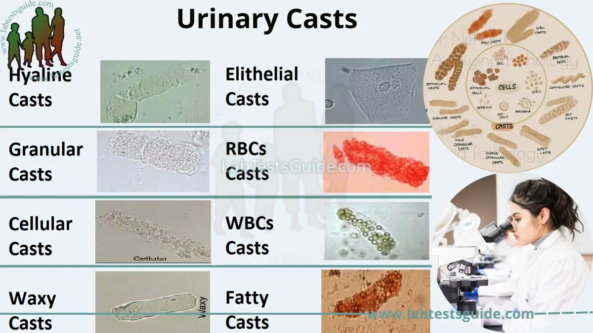
Hyaline Casts
Composed purely of Tamm-Horsfall (uromodulin) protein. Low refractive index; hard to spot.
RBC Casts
Red blood cells trapped within matrix. Indicates bleeding of glomerular origin.
WBC Casts
White blood cells embedded in matrix. Distinguishes upper from lower UTI.
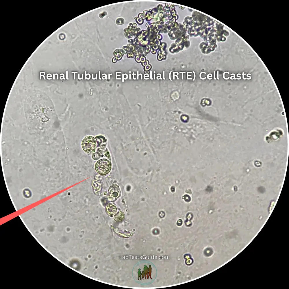
RTE Cell Casts
Desquamated tubular epithelial cells. Indicates extensive necrosis and sloughing of renal nephron lining.
Epithelial Casts
Renal tubular epithelial (RTE) cells sloughed off into matrix due to severe tubular injury.
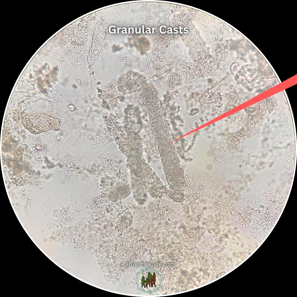
Granular Casts
Degenerated cellular casts. Coarse or fine texture depending on stasis duration.
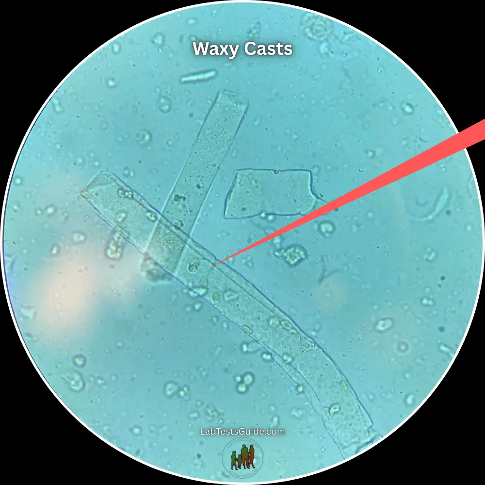
Waxy Casts
Ultimate stage of cast degeneration. High refractive index with cracked margins.
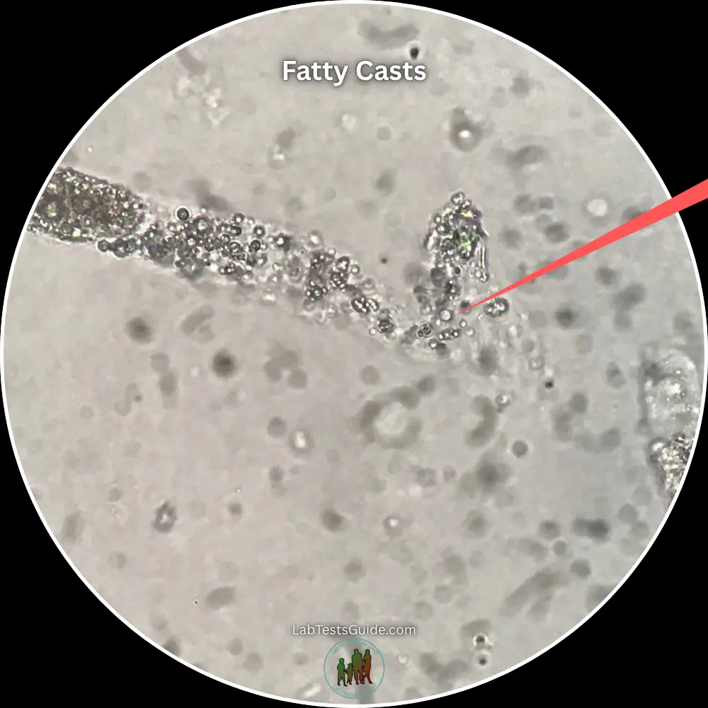
Fatty Casts
Contains lipid droplets or oval fat bodies. Polarized light reveals pathognomonic "Maltese cross" figures.
Broad Casts
Markedly wider diameter formed in dilated collecting ducts; indicates extensive nephron loss and atrophy.
Pigmented Casts
Matrix impregnated with endogenous pigments like hemoglobin, myoglobin, or bilirubin.
Mixed Cellular Casts
Contains two or more distinct cell lines (e.g., RBCs + WBCs or RBCs + RTE cells) in a single matrix.
Cylindroid Casts
Cast-like structures featuring one classic parallel cylindrical border and one long, tapered, ribbon-like tail.
Microscopic & Clinical Comparison
Comprehensive laboratory evaluation of urinary cast morphologies, cellular markers, and underlying pathology.
| Cast Type | Microscopic Morphology | Clinical Significance | Associated Pathologies / Causes |
|---|---|---|---|
Hyaline Casts
Physiological | Colorless, homogeneous, transparent with a low refractive index and rounded ends. Composed solely of uromodulin. | Usually non-pathological; indicates urinary stasis, concentrated urine, or transient renal hemodynamic changes. | Dehydration, strenuous physical exercise, fever, emotional stress, loop diuretic therapy. |
RBC Casts
Glomerular | Erythrocytes embedded within matrix; yellow-orange to reddish-brown hue. May undergo lysis into hemoglobin casts. | Diagnostic hallmark of intrinsic renal disease with severe basement membrane rupture in glomeruli. | Post-streptococcal glomerulonephritis, IgA nephropathy, Lupus nephritis, Goodpasture syndrome, ANCA vasculitis. |
WBC Casts
Infectious / Inflammatory | Polymorphonuclear neutrophils densely packed in protein matrix; distinct lobed nuclei and granular cytoplasm. | Key marker differentiating upper renal tract infection/inflammation from lower urinary tract infection (cystitis). | Acute pyelonephritis, tubulointerstitial nephritis, renal abscess, transplant rejection. |
RTE Cell Casts
Tubular Injury | Renal tubular epithelial cells arranged in rows or randomly attached; large centrally placed round nuclei. | Indicates acute sloughing and destruction of nephron tubular epithelium due to ischemic or toxic insult. | Acute Tubular Necrosis (ATN), heavy metal poisoning (lead/mercury), ethylene glycol toxicity, nephrotoxic drugs (aminoglycosides). |
Granular Casts
Degenerative | Coarse or fine granules dispersed throughout matrix; derived from breakdown of cellular casts or protein aggregates. | Reflects prolonged urinary stasis within nephrons and progressive breakdown of cellular debris. | Advanced ATN ("muddy brown" granular casts), chronic kidney disease, severe dehydration, pyelonephritis. |
Waxy Casts
End-Stage | High refractive index, yellowish opaque appearance, smooth homogenous texture with sharp, notched, or cracked ends. | Final stage of cellular/granular cast evolution; represents extreme, prolonged renal stasis and severe tubular atrophy. | Advanced Chronic Kidney Disease (CKD), End-Stage Renal Disease (ESRD), diabetic nephropathy, severe renal allograft rejection. |
Fatty Casts
Lipiduria | Matrix contains highly refractive lipid droplets or oval fat bodies. Exhibits polarized "Maltese cross" anisotropy. | Signifies severe glomerular capillary permeability breakdown leading to heavy filtration of lipoproteins. | Nephrotic Syndrome, minimal change disease, focal segmental glomerulosclerosis (FSGS), membranous nephropathy. |
Pigmented Casts
Metabolic | Distinct color saturation: deep red-brown (hemoglobin/myoglobin), golden-brown (bilirubin), or dark brown (methemoglobin). | Indicates filtration of massive endogenous metabolic pigment loads causing direct tubular oxidative toxicity. | Rhabdomyolysis (myoglobinuria), intravascular hemolysis (hemoglobinuria), severe obstructive jaundice (bilirubinuria). |
Introduction to Urinary Casts
What Are Urinary Casts?
Urinary casts are cylindrical, microscopic structures formed exclusively within the lumens of distal convoluted tubules and collecting ducts of the nephron. They feature a core matrix composed of Tamm-Horsfall glycoprotein (uromodulin) secreted by renal tubular cells, which can entrap cells, debris, or lipids during formation.
Why Urinary Casts Matter
Because casts are shaped by the tubular lumen itself, their presence directly confirms that detected cellular or chemical abnormalities originate directly from the kidney parenchyma—not from lower urinary tract structures like the ureters, bladder, or urethra.
Overview of Their Role in Kidney Health
In clinical urinalysis, cast identification serves as a crucial non-invasive diagnostic tool. By analyzing cast morphology and cellular composition, clinicians can localize intrarenal disease, monitor acute kidney injury progression, differentiate upper vs. lower urinary tract pathology, and assess overall nephron degradation.
Key Takeaways & Quick Reference
Quick Facts
- Site of Origin: Distal convoluted tubules & collecting ducts.
- Matrix Core: Tamm-Horsfall protein (uromodulin).
- Diagnostic Role: Localizes pathology directly to kidney parenchyma.
- Microscopy: Evaluated under Low Power Field (LPF) for counting.
Key Takeaways
- Casts confirm intrarenal origin, excluding lower UTI/bleeding.
- Cellular casts degenerate: Cellular → Granular → Waxy.
- RBC casts are pathognomonic for active glomerulonephritis.
- WBC casts differentiate acute pyelonephritis from lower cystitis.
When Casts Are Normal
Small amounts of Hyaline Casts (0–2 / LPF) are considered normal baseline findings. They form during temporary urinary stasis or increased protein concentration without structural kidney damage.
When Casts Suggest Disease
The presence of Cellular, Broad, or Waxy Casts indicates ongoing nephron injury, active inflammatory/infectious processes, or severe structural disruption of the renal filtration barrier.
What Are Urinary Casts?
Mechanisms of intranephronic cast formation, anatomical localization, and structural matrix composition.
Definition
Urinary casts are microscopic, cylindrical structures created through the gelation of Tamm-Horsfall protein within renal tubules. Because they form exclusively inside the nephron lumen, they take on the exact shape and size of the tubule and serve as direct biological molds of intrarenal conditions.
Where Casts Form
Casts precipitate specifically in the distal convoluted tubules (DCT) and collecting ducts. These distal nephron sites provide the necessary physiological conditions for protein aggregation: high solute concentration, low flow velocity, acidic pH, and high sodium content.
Formation Process (Gelation Kinetics)
Composition of Urinary Casts
How Urinary Casts Form
Key factors, biochemical conditions, and tubular dynamics that drive cast matrix precipitation.
Role of Tamm-Horsfall Protein (Uromodulin)
Uromodulin is a high-molecular-weight glycoprotein secreted exclusively by epithelial cells lining the thick ascending limb of Henle's loop. Under favorable conditions, monomeric uromodulin undergoes polymerization, forming a dense, gel-like fibrillar network that serves as the essential matrix backbone for all urinary casts.
Conditions Favoring Formation
- High Solute Load: Elevated urinary ionic strength promotes protein aggregation.
- Presence of Free Light Chains: Bence-Jones proteins accelerate gelation kinetics.
- High Albumin Concentration: Increased glomerular permeability adds matrix protein.
Factors Affecting Production Rate
- Tubular Health: Intact distal tubule epithelia ensure adequate uromodulin secretion.
- Glomerular Filtration Rate: Reduced GFR increases contact time in tubules.
- Epithelial Sloughing: Cell death provides particulate matter to entrap.
Critical Environmental Drivers
Acidic pH (< 6.0) dramatically decreases the solubility of uromodulin, causing rapid aggregation. Simultaneously, concentrated urine (high specific gravity / hypertonicity) forces protein molecules into close proximity, accelerating gelation.
Sluggish tubular flow (due to dehydration, oliguria, or renal artery stenosis) provides the necessary dwell time for polymerized uromodulin fibrils to entangle and entrap passing cells or debris into a cohesive mold.
10 Types of Urinary Casts (Comprehensive Guide)
Detailed breakdown of cast morphology, underlying etiologies, and diagnostic significance across major clinical presentations.
Hyaline Casts
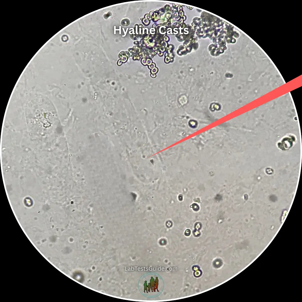
Description: Composed almost exclusively of Tamm-Horsfall protein (uromodulin). Smooth, transparent, homogeneous cylindrical structures with a low refractive index.
Causes: Dehydration, strenuous physical exertion, high fever, or systemic stress.
Clinical Significance: Frequently non-pathological (0–2/LPF normal). High numbers indicate prerenal azotemia or reduced flow.
Microscopic Appearance: Colorless and faint; difficult to visualize under standard brightfield microscopy without reducing illumination.
Associated Conditions: Dehydration, congestive heart failure, prerenal AKI.
Red Blood Cell (RBC) Casts
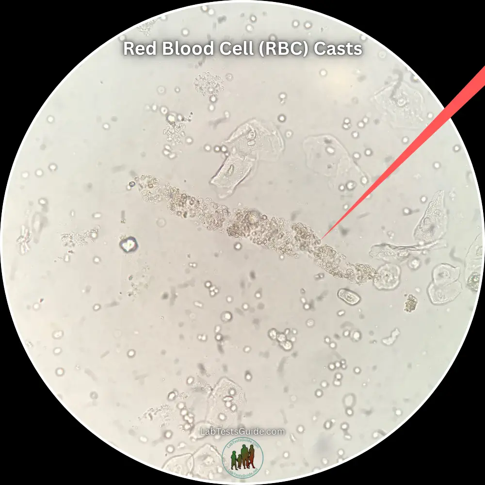
Description: Protein matrix embedded with intact or dysmorphic erythrocytes, giving a yellowish-orange to brown hue.
Causes: Disruption of the glomerular filtration barrier allowing red blood cells into the tubule lumen.
Diseases Associated: Post-streptococcal glomerulonephritis, IgA nephropathy, Lupus nephritis, Goodpasture syndrome, ANCA vasculitis.
Clinical Importance: Diagnostic hallmark for active glomerular hemorrhage (nephritic syndrome).
White Blood Cell (WBC) Casts
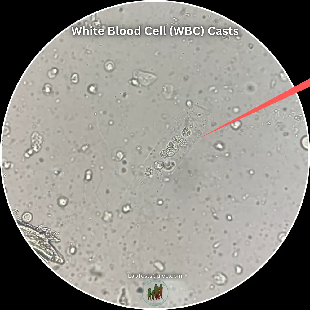
Description: Casts densely packed with leukocytes (predominantly neutrophils), showing visible nuclear detail.
Causes: Active interstitial inflammation or infection within the renal parenchyma.
Kidney Infections: Acute pyelonephritis (differentiates upper UTI from uncomplicated lower cystitis).
Autoimmune Diseases: Acute interstitial nephritis (AIN), active lupus nephritis, acute renal transplant rejection.
Renal Tubular Epithelial (RTE) Cell Casts
Description: Cylindrical structures containing sloughed rectangular or round tubular epithelial cells.
Causes: Direct toxic or ischemic injury to tubular epithelial lining causing necrosis and detachment.
Clinical Importance: Strong indicator of Acute Tubular Necrosis (ATN), heavy metal toxicity, viral nephropathy, or drug-induced nephrotoxicity.
Granular Casts
Fine Granular Casts: Contain small, uniform particles from late-stage cellular breakdown or protein aggregation.
Coarse Granular Casts: Feature larger, irregular granules representing early tubular cell degeneration.
Clinical Significance: Classic "muddy brown" coarse granular casts strongly indicate Acute Tubular Necrosis (ATN).
Waxy Casts
Description: Highly refractive, rigid, yellow or translucent casts with distinct notched edges, cracks, and square ends.
Causes: Severe prolonged urinary stasis within dilated tubules allowing advanced protein degradation.
Advanced Kidney Disease: Signifies chronic kidney disease (CKD), end-stage renal disease (ESRD), or severe renal stasis.
Fatty Casts
Description: Casts containing intracellular or extracellular lipid droplets, triglycerides, and oval fat bodies.
Maltese Cross Appearance: Displays characteristic birefringent "Maltese cross" pattern under polarized light.
Nephrotic Syndrome: Pathognomonic for heavy proteinuria and lipiduria seen in Nephrotic Syndrome, diabetic nephropathy, or FSGS.
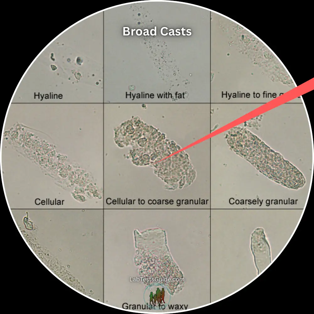
Broad Casts
Description: Abnormally wide casts (2–6x standard cast width) formed in dilated collecting ducts.
End-Stage Kidney Disease: Termed "renal failure casts," reflecting compensatory dilation of surviving nephrons.
Clinical Importance: Indicates widespread loss of functional nephron mass and severe renal insufficiency.
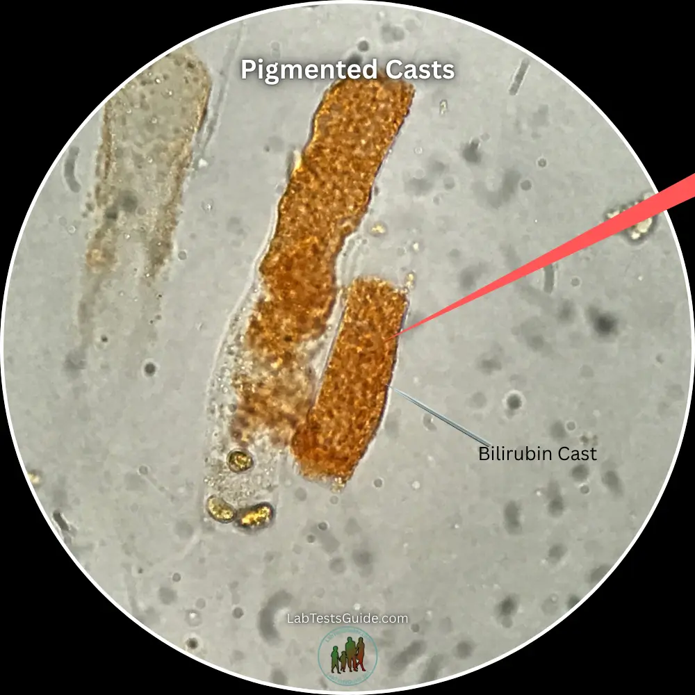
Pigmented Casts
Hemoglobin Casts: Reddish-brown casts resulting from filtered free hemoglobin during intravascular hemolysis.
Myoglobin Casts: Dark brown or tea-colored casts from massive muscle necrosis (rhabdomyolysis).
Bilirubin Casts: Distinct deep yellow-orange casts in severe conjugated hyperbilirubinemia or obstructive jaundice.
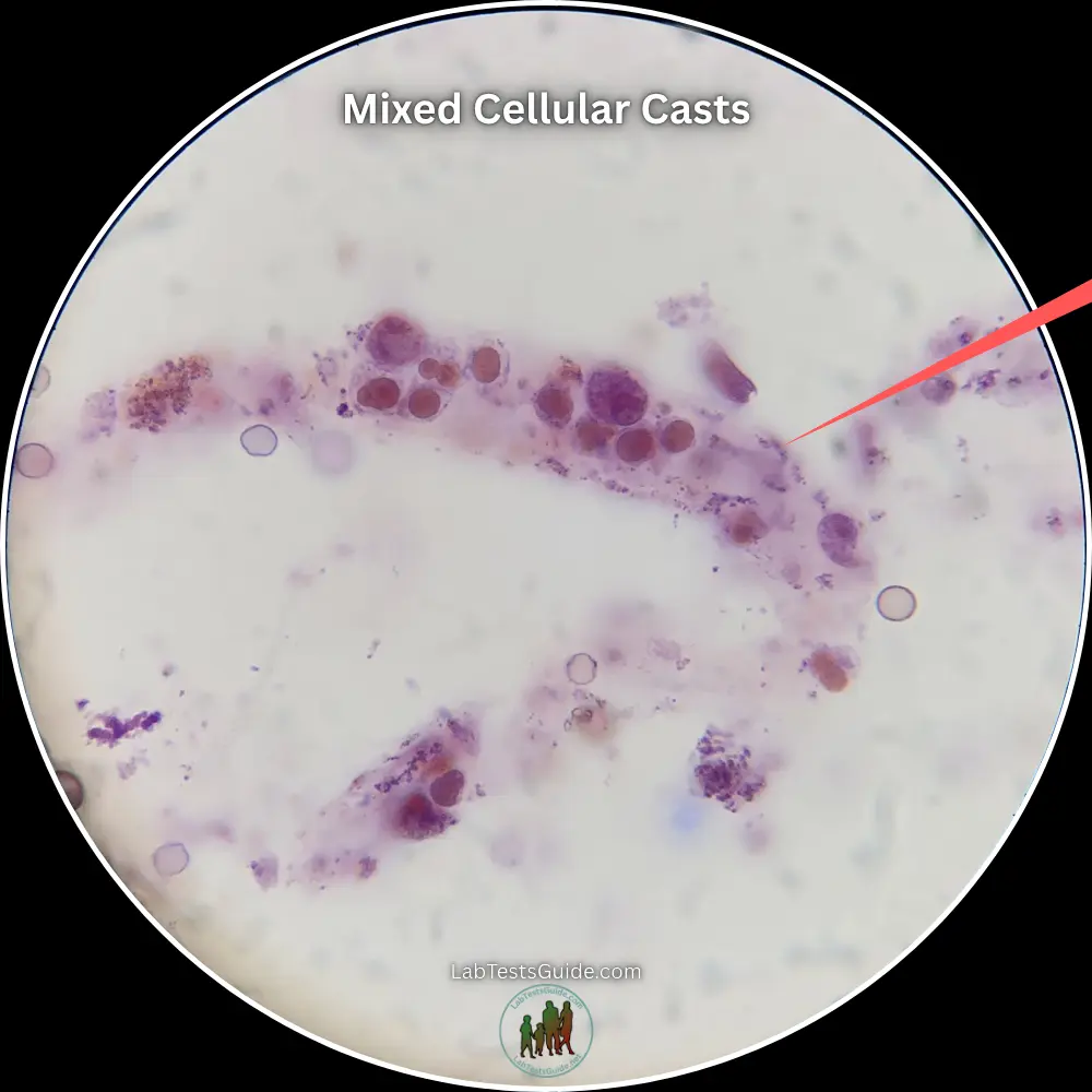
Mixed Cellular Casts
Definition: Casts containing two or more distinct cell populations (e.g., RBCs + WBCs, or WBCs + RTE cells) in one matrix.
Clinical Relevance: Points to complex or multifactorial pathology, such as crescentic glomerulonephritis combined with acute tubular damage.
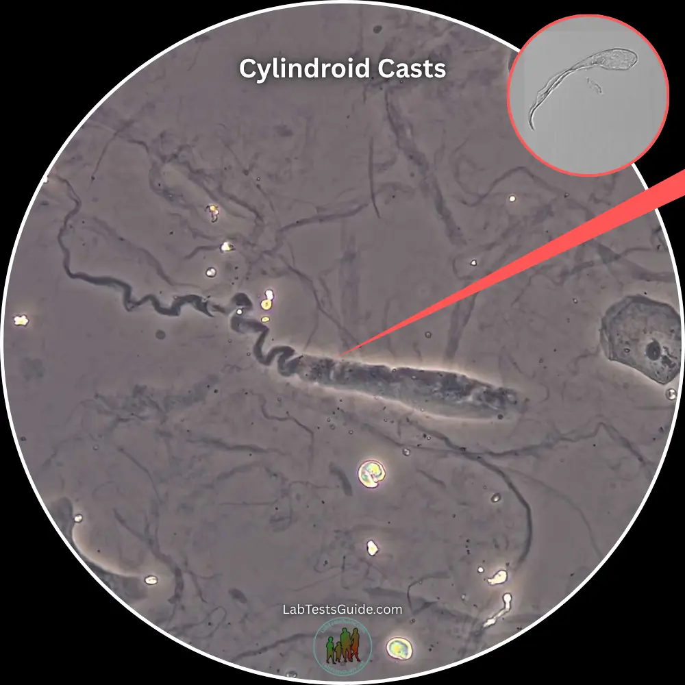
Cylindroid Casts
Description: Hybrid structures resembling hyaline or granular casts at one end while tapering into a fine strand or tail at the opposite end.
Pathophysiology: Formed when uromodulin (Tamm-Horsfall protein) gelation occurs under partial or incomplete tubular flow, preventing full cylindrical molding.
Clinical Importance: Carries the exact same diagnostic implications as standard hyaline or granular casts; the tapered morphology has no separate pathologic meaning.
Urinary Casts Classification
Taxonomic grouping based on internal architecture, cellular presence, and stage of protein or organelle degeneration.
1. Cellular Casts
Casts containing identifiable intact cellular elements entrapped within the Tamm-Horsfall matrix. Their presence directly localizes severe cell injury, bleeding, or leukocyte migration to the renal parenchyma.
2. Acellular Casts
Casts composed purely of protein matrix, lipid inclusions, or metabolic/pigment deposits without intact cellular structures.
3. Degenerative Casts
Represents the progressive morphological breakdown sequence as cellular casts decompose during prolonged tubular transit time.
Freshly entrapped intact RBCs, WBCs, or RTE cells with clear borders.
Cells lyse, fragmenting into coarse plasma-membrane granules.
Granules break down into tiny, homogeneous particulate matter.
Homogenized, rigid, brittle matrix indicating prolonged urinary stasis.
Normal vs. Abnormal Urinary Casts
Clinical thresholding to differentiate transient physiological findings from active renal parenchymal disease.
Which Casts Can Be Normal?
Occasional cast formation can occur in healthy individuals due to temporary hemoconcentration, mild urinary stasis, or transient alterations in renal hemodynamics without underlying structural kidney damage.
Composed entirely of uromodulin. Frequently seen following strenuous exercise, high fever, severe dehydration, or exposure to cold environments.
Can occasionally appear in very low numbers after extreme physical exertion due to temporary protein aggregation without tubule degeneration.
Which Casts Always Indicate Disease?
Certain cast morphologies reflect active renal cell necrosis, inflammatory filtration barrier damage, or severe intrarenal stasis, and are never considered normal baseline findings.
Confirm active glomerular bleeding (Glomerulonephritis) or parenchymal inflammation (Pyelonephritis / AIN).
Signify active desquamation and ischemic or nephrotoxic injury to tubular cells (Acute Tubular Necrosis).
Pathognomonic for advanced chronic kidney disease (CKD), end-stage renal disease (ESRD), or heavy lipiduria in Nephrotic Syndrome.
| Feature | Normal / Physiological Findings | Abnormal / Pathological Findings |
|---|---|---|
| Dominant Cast Types | Hyaline, rare fine granular | RBC, WBC, RTE, Coarse Granular, Waxy, Fatty, Broad |
| Quantification | Isolated (0–2 / LPF) | Persistent or moderate-to-high counts (> 2–5 / LPF) |
| Primary Mechanism | Concentrated urine + low flow rate | Glomerular barrier rupture, tubular necrosis, nephron dilation |
| Clinical Action Required | Rehydration & clinical observation | Urgent nephrology evaluation, diagnostic workup & intervention |
Causes of Urinary Casts
Comprehensive breakdown of physiological stressors, primary renal pathologies, and systemic conditions driving cast formation.
1. Physiological Triggers
Produces low urinary volume and high solute concentration, promoting uromodulin polymerization into hyaline casts.
Transient renal vasoconstriction lowers GFR, causing temporary urinary stasis and hyaline or fine granular cast formation.
Systemic hyperthermic metabolic stress increases protein excretion and urinary concentration, temporarily raising cast output.
2. Primary Kidney Diseases
Ischemic or toxic tubular insult leads to rapid epithelial cell necrosis, desquamation, and classic "muddy brown" granular cast formation.
Long-term nephron loss causes compensatory hypertrophy and dilation of remaining tubules, yielding abnormally wide, rigid waxy casts.
Inflammatory rupture of the capillary basement membrane allows red blood cells to enter the tubules, forming pathognomonic RBC casts.
Acute bacterial invasion of the renal parenchyma causes neutrophil migration into tubules, generating dense WBC casts.
Severe glomerular podocyte effacement causes massive proteinuria and hyperlipidemia; tubular reabsorption of filtered lipids forms fatty casts showing a "Maltese cross" under polarized light.
3. Systemic Conditions
4. Drug-Induced Injury
Exogenous nephrotoxins directly injure renal tubular epithelial cells or induce hypersensitivity reactions within the tubulointerstitium.
Diseases Associated with Different Cast Types
Clinical mapping linking specific urinary sediment morphologies to localized renal lesions and underlying systemic pathology.
| Cast Type | Primary Disease Associations | Pathophysiologic Mechanism |
|---|---|---|
| RBC Casts Glomerular |
| Gaps in the glomerular basement membrane allow erythrocyte transit into Bowman's space and tubular matrix trapping. |
| WBC Casts Tubulointerstitial |
| Exudation of polymorphonuclear leukocytes across the interstitial capillary border directly into renal tubules. |
| RTE Cell Casts Tubular Epithelial |
| Direct injury to proximal/distal tubular cells leading to detachment, necrosis, and entrapment in uromodulin. |
| Fatty Casts Glomerular / Lipid |
| Massive proteinuria causes renal tubule endocytosis of filtered lipoproteins; decaying cells spill intracellular cholesterol. |
| Waxy & Broad Casts Terminal Stasis |
| Extreme intrarenal urinary stasis in severely dilated or hypertrophic surviving tubules allowing complete matrix degeneration. |
Diagnostic Urinalysis Patterns
Classic multi-variable sediment profiles used to differentiate acute renal syndromes at the bedside.
Active Glomerulonephritis
Heavy Proteinuria / Glomerulopathy
Acute Tubular Necrosis
Pyelonephritis vs. AIN
Symptoms Associated with Urinary Casts
While urinary casts are microscopic findings, their presence frequently aligns with specific localized renal, lower urinary tract, or systemic manifestations.
1. Kidney Disease Symptoms
Direct manifestations of renal parenchymal injury, altered glomerular hemodynamics, and impaired fluid homeostasis.
2. Urinary Symptoms
Gross alterations in urine physical characteristics, output volumes, and micturition mechanics.
3. Systemic Symptoms
Widespread physiological signs accompanying underlying inflammatory, infectious, autoimmune, or uremic pathologies.
Common in upper tract infections (Acute Pyelonephritis) or hypersensitivity-driven Drug-Induced Acute Interstitial Nephritis.
General weakness, loss of appetite, nausea, and metallic taste secondary to accumulation of nitrogenous waste products in AKI or CKD.
Joint pain and systemic skin manifestations typical of underlying autoimmune vasculitis or Systemic Lupus Erythematosus (Lupus Nephritis).
How Urinary Casts Are Detected
Standardized clinical workflows for pre-analytical collection, specimen processing, sediment concentration, and microscopic identification.
1. Sample Collection Protocol
Optimizing sample integrity is critical to prevent in vitro lysis or dissolution of delicate mucoprotein casts prior to examination.
2. Laboratory Processing Procedure
Standardized physical concentration of urinary elements to yield a representative sediment button.
3. Urine Sediment Microscopy & Evaluation
Microscopic identification requires systematic optics adjustments to distinguish low-refractive-index casts from surrounding background artifact.
Used to scan the edges of the coverslip where casts naturally migrate due to capillary action. Quantified as average number per LPF (e.g., 2–5 casts / LPF).
Utilized for detailed morphological identification—classifying internal matrix contents as intact cellular lines, granular debris, lipids, or waxy notches.
Phase-Contrast Optics: Enhances low-refractive hyaline matrices.
Polarized Light: Identifies anisotropic lipid drops in fatty casts ("Maltese Cross").
Microscopic Appearance of Urinary Casts
Key structural features for accurate cast identification, common visual artifacts, and differential diagnostic criteria under light microscopy.
1. Key Identification Features
True urinary casts share fundamental structural hallmarks reflecting their cylindrical molding within the distal convoluted tubule and collecting duct.
2. Common Microscopic Pitfalls
Extrainfectious or environmental contaminants frequently mimic cast morphology, risking false-positive renal parenchymal pathology reports.
3. Differential Diagnosis Matrix
Differential features to distinguish true casts from pseudocasts and differentiate cell types within intact cellular casts.
| Microscopic Feature | True Urinary Cast | Pseudocast / Artifact | Differential Diagnostic Significance |
|---|---|---|---|
| Borders & Termini | Parallel edges, blunt/rounded ends | Frayed, tapered, or irregular ends | Distinguishes nephron-molded matrix from mucus/fibers. |
| Matrix Inclusions | Cells/granules enclosed inside matrix | Elements resting on top or around debris | Confirms intrarenal origin rather than bladder/voiding clump. |
| RBC vs. WBC Cast | Yellow-orange hue, dysmorphic RBCs | Multilobed nuclei, larger granular cells | Glomerulonephritis (RBC) vs. Pyelonephritis/AIN (WBC). |
| WBC vs. RTE Cell Cast | Neutrophils with segmented nuclei | Large round cells, single eccentric nucleus | Tubulointerstitial inflammation vs. Direct ATN necrosis. |
| Hyaline vs. Waxy Cast | Low refraction, smooth transparent matrix | High refraction, yellowish, cracked/notched edges | Transient physiological stasis vs. Severe chronic renal failure. |
Urinary Cast Interpretation
A systematic framework for evaluating urinary sediment, correlating microscopic cast morphology with renal disease states, and reporting standardized findings.
1. Step-by-Step Microscopic Interpretation
Follow this 4-step sequence during urinary sediment examination to systematically identify and classify cast structures.
Artifact Exclusion
Differentiate true casts from pseudocasts, fibers, or mucus threads by confirming parallel sides and a rounded matrix border under 10× LPF.
Matrix Evaluation
Determine refractive index and texture under 40× HPF: clear/transparent (Hyaline), granular/particulate, or broad/brittle with notches (Waxy).
Inclusion Analysis
Identify entrapped cellular elements (RBCs, WBCs, RTE cells), bacterial clusters, or oval fat bodies embedded within the protein gel.
Width & Frequency
Quantify average count per Low Power Field (LPF) and assess cast width (standard vs. broad casts indicative of collecting duct dilation).
2. Clinical & Urinalysis Correlation
Correlate cast findings with dipstick parameters and laboratory indicators to localize the renal pathology.
RBC Casts + Positive Blood Dipstick + Dysmorphic Red Cells + Mild/Moderate Proteinuria.
Fatty & Hyaline Casts + 4+ Dipstick Proteinuria + Oval Fat Bodies + Maltese Cross under polarized light.
"Muddy Brown" Coarse Granular & RTE Casts + Free RTE Cells + Low Urine Specific Gravity (≤1.010).
WBC Casts + Positive Leukocyte Esterase & Nitrite + Pyuria (WBC clumps) ± Urinary Eosinophils.
3. Reporting Guidelines
Standardized semi-quantitative terminology for clinical urinalysis reporting based on 10× Low Power Field (LPF) enumeration.
| Reporting Term | Hyaline Casts (per LPF) | Pathological Casts (per LPF) |
|---|---|---|
| None Seen | 0 / LPF | 0 / LPF |
| Rare / Occasional | 1 – 2 / LPF | 1 / LPF |
| Few (1+) | 3 – 5 / LPF | 2 – 5 / LPF |
| Moderate (2+) | 6 – 10 / LPF | 6 – 10 / LPF |
| Many / Numerous (3+) | > 10 / LPF | > 10 / LPF |
Normal Reference Range & Reporting Standards
Establishing physiological baselines for urinary cast excretion, healthy sediment findings, and standardized clinical laboratory reporting conventions.
1. Normal Reference Ranges
Quantification thresholds per Low Power Field (LPF) under standard urine sediment concentration (10:1 ratio, 400 × g).
2. Non-Pathological "Normal" Findings
Transient cast excretion can occur in healthy individuals due to temporary intrarenal hemoconcentration or physiological stress.
Temporary athletic proteinuria and reduced renal perfusion trigger a transient surge of hyaline and fine granular casts, resolving within 24–48 hours of rest.
High urine specific gravity (>1.025) and acidic pH promote uromodulin (Tamm-Horsfall) gelation, causing isolated hyaline cast precipitation without tubular damage.
Rapid fluid shifts or febrile states induce transient sluggish tubular flow and physiological hyaline cast formation in the distal nephron segment.
3. Standardized Reporting Conventions
Laboratory protocols for semi-quantitative grading and narrative modifiers to ensure clear communication with ordering clinicians.
Semi-Quantitative Enumeration Scale
- Rare / Occasional: Average of 1 cast per LPF or seen only in multiple fields.
- Few (1+): 2 to 5 casts per Low Power Field (10×).
- Moderate (2+): 6 to 10 casts per Low Power Field (10×).
- Many / Numerous (3+): Greater than 10 casts per Low Power Field (10×).
Mandatory Descriptive Modifiers
- Matrix Width Annotation: Must explicitly designate "Broad Casts" when width exceeds 3× standard tubule diameter.
- Pigmentation / Coloration: Record internal staining (e.g., "Bilirubin-stained", "Hemoglobin-pigmented", or "Muddy Brown").
- Mixed Morphologies: Report dominant matrix type alongside specific inclusion lines (e.g., "Hyaline cast with 20% RTE cell inclusion").
Clinical Significance & Diagnostic Importance
Correlating individual urinary cast morphologies with primary renal lesion sites, disease severity, and rapid diagnostic decision drivers.
1. Detailed Clinical Significance by Cast Type
Urinary casts serve as "nephron biopsies" providing direct evidence of tubular status, glomerular capillary wall integrity, or interstitial inflammation.
Occur normally in low numbers. High counts indicate concentrated urine, high fever, intense exertion, congestive heart failure, or loop diuretic use without direct tubular injury.
Pathognomonic for active glomerulonephritis (e.g., IgA Nephropathy, Lupus Nephritis, ANCA Vasculitis). Indicates mechanical breach of the basement membrane and intra-tubular trapped blood.
Differentiates upper UTI (Acute Pyelonephritis) from lower UTI (Cystitis). Also seen in Acute Interstitial Nephritis (AIN) and renal transplant rejection.
Key hallmark of ischemic or nephrotoxic Acute Tubular Necrosis (ATN), viral infections (CMV), interstitial nephritis, and heavy metal or drug intoxication (e.g., Aminoglycosides).
Represents degenerated cell inclusions or aggregated serum proteins. "Muddy Brown" coarse granular casts are diagnostic for ischemic/toxic ATN in acute kidney injury (AKI).
Indicates extreme urinary stasis in dilated, hypertrophied, or severely damaged collecting ducts. Characteristic of Chronic Kidney Disease (CKD Stage 4/5) and severe end-stage renal graft failure.
2. Quick Interpretation Guide
Rapid visual correlation matrix matching primary urine sediment cast findings with clinical syndromes.
| Primary Cast Identified | Predominant Site | Suspected Diagnosis |
|---|---|---|
| RBC Casts | Glomerulus | Glomerulonephritis |
| WBC Casts | Interstitium / Tubule | Pyelonephritis / AIN |
| RTE / Coarse Granular | Tubular Epithelium | Acute Tubular Necrosis |
| Fatty Casts | Glomerular Barrier | Nephrotic Syndrome |
| Broad Waxy Casts | Collecting Duct | Advanced CKD / ESRD |
3. Diagnostic Importance & Utility
Why cast analysis remains a crucial non-invasive bedside tool in modern nephrology.
Differentiates intrarenal disease (e.g., intrinsic AKI due to glomerulonephritis or ATN) from prerenal azotemia and postrenal obstructive uropathies.
Detection of RBC casts or dysmorphic red cell profiles serves as a primary indication to order an urgent percutaneous renal biopsy.
Serial quantification of cellular casts tracks disease resolution or flare-ups in autoimmune conditions such as Systemic Lupus Erythematosus (SLE).
When to See a Doctor, Warning Signs & Emergency Symptoms
Establishing patient-facing and clinical red-flag criteria to guide timely medical evaluation, nephrology referral, and emergency intervention based on urinalysis cast findings.
1. When to Schedule an Evaluation
Non-emergent situations warranting routine primary care follow-up or scheduled nephrology consultation.
Persistent presence of >2 hyaline or fine granular casts per field on repeat routine urinalysis without underlying physical exertion or acute illness.
Routine dipstick positive for trace blood or microscopic RBCs without dysuria or overt urinary tract infection signs.
Slight changes in urine consistency suggesting low-grade proteinuria, especially in patients with diabetes or hypertension.
2. Warning Signs (Urgent Care)
Signs of active parenchymal inflammation or progressive renal injury requiring prompt medical workup within days.
Identification of RBC casts, WBC casts, or RTE cell casts on microscopy, indicating active glomerulonephritis, pyelonephritis, or ATN.
Swelling in the lower extremities, ankles, or periorbital region paired with fatty casts or nephrotic-range proteinuria.
Fever, flank pain, malar rash, or joint pain paired with cellular casts (suggestive of Lupus Nephritis or Vasculitis).
3. Emergency Red-Flag Symptoms
Life-threatening clinical manifestations requiring immediate emergency room admission to manage acute kidney injury (AKI), uremia, or severe fluid overload.
Sudden cessation of urine output or producing less than 400 mL/day alongside broad, waxy, or "muddy brown" coarse granular casts.
Visible tea-colored, cola-colored, or frankly bloody urine accompanied by RBC casts and rapidly worsening kidney function.
Shortness of breath, confusion, severe nausea/vomiting, pericardial friction rub, or refractory hypertension indicative of end-stage uremic toxicity.
Artifacts vs. Casts: Differential Diagnosis & Look-Alikes
Distinguishing true proteinaceous renal casts from common microscopic contaminants, pseudocasts, and structural artifacts in urine sediment.
Misidentifying non-cast structures as true renal casts is a primary source of laboratory error in routine urinalysis. True casts possess parallel sides, rounded ends, and a matrix composed of gelled Tamm-Horsfall protein. In contrast, artifacts lack a true protein matrix and often exhibit irregular, tapered, or refractive features.
| Microscopic Element | Key Visual Characteristics | Distinguishing Feature from True Casts | Clinical Significance |
|---|---|---|---|
| Mucus Threads | Long, thin, ribbon-like strands with faint, wavy longitudinal striations and frayed ends. | Lacks distinct parallel sides and rounded borders; threads intertwine rather than forming distinct cylindrical blocks. | Benign; indicates lower tract irritation or normal urethral/vaginal contamination. |
| Synthetic / Clothing Fibers | Flat or thick strands with high refractivity, sharp split ends, and variable color tinting. | Extremely birefringent under polarized light; much larger and denser than renal tubules. | Extrinsic artifact (clothing, lint, paper towels); zero clinical relevance. |
| Hair Shafts | Thick, uniform cylinders with a visible central medulla and distinct outer cuticle scales. | Opaque center with definitive structural anatomy; far larger than any renal tubule. | Extrinsic specimen contamination. |
| Crystal Aggregates (Pseudocasts) | Clumped urates, phosphates, or calcium oxalate crystals forming oblong shapes. | Lacks a gelled mucoprotein backbone; edges are jagged, crystalline, and dissolve with specific reagents (pH change). | Hypercrystalluria; can obscure true cellular elements if abundant. |
| Glass Scratches / Air Bubbles | Perfectly round rings with dark borders (bubbles) or fixed linear slide defects. | Remains stationary when urine sediment shifts; optical plane artifact. | Equipment artifact. |
| Bacterial Aggregates | Dense clusters of motile or non-motile bacteria forming elongated masses. | Lacks matrix borders; granular look is composed entirely of distinct, uniform microbes rather than degenerated protein/cells. | Bacteriuria/UTI; mistaking for granular casts leads to misdiagnosing intrinsic renal involvement. |
Rule of Matrix & Borders
True casts always show a continuous protein matrix holding the structures together, along with uniform cylindrical dimensions that match the renal tubule diameter.
Polarized Light Verification
Synthetic fibers (e.g., cotton, nylon) show strong birefringence under polarized light, whereas true hyaline and granular casts do not (except for fatty casts showing Maltese crosses).
Clinical Diagnostic Algorithm for Urinary Cast Evaluation
A structured, step-by-step decision pathway for clinicians and laboratory professionals from initial sediment detection to diagnostic triage and targeted management.
Specimen Screening & Screening Verification
Low Power (10x)Scan freshly centrifuged urine sediment under Low Power Field (100x magnification) near the edges of the coverslip. Confirm presence of parallel-sided cylindrical protein matrices.
Morphological Classification & Inclusion Analysis
High Power (40x)Identify specific inclusions within the uromodulin matrix under High Power Field (400x magnification) or Phase-Contrast Microscopy.
Hyaline Casts (0–2/LPF normal; >2 suggests pre-renal stasis, concentrated urine, or exertion).
RBC Casts → Active Glomerular Bleeding (Glomerulonephritis, IgA Nephropathy, Vasculitis).
WBC Casts → Tubulointerstitial Inflammation / Infection (Pyelonephritis, AIN).
RTE Casts / Muddy Brown Casts → Tubular Injury (Ischemic / Toxic ATN).
Fatty Casts → Severe Proteinuria / Glomerular Permeability (Nephrotic Syndrome).
Waxy / Broad Casts → Severe Intrarenal Stasis / Advanced Nephron Loss (CKD Stage 4/5, ESRD).
Correlation with Dipstick Chemistry & Serology
Lab CorrelationCross-reference microscopic findings with reagent strip urinalysis and routine serum metabolic panels:
- RBC Casts + Positive Dipstick Blood + Proteinuria: Glomerular Syndrome. Check ANA, ANCA, anti-GBO, C3/C4, ASO titers.
- WBC Casts + Leukocyte Esterase + Nitrite: Acute Pyelonephritis. Order Urine Culture & Sensitivity.
- WBC Casts + Eosinophiluria + Negative Nitrite: Drug-Induced Acute Interstitial Nephritis (AIN). Review drug list (NSAIDs, PPIs, Antibiotics).
- RTE / Granular Casts + Elevated Cr/BUN: Acute Kidney Injury. Calculate FENa/FEUrea to differentiate Prerenal vs. Intrinsic ATN.
- Fatty Casts + Heavy Proteinuria (3+/4+): Nephrotic Syndrome. Order 24-hr Urine Protein / UPCR and Serum Albumin.
Clinical Action & Triage Pathway
ManagementReassure patient, optimize oral hydration, and recheck routine urinalysis in 2–4 weeks if clinically indicated.
Initiate targeted antibiotic therapy (Pyelonephritis), withdraw offending nephrotoxin (AIN/ATN), and monitor serum creatinine every 24–48 hours.
Urgent Nephrology Consultation. Schedule renal ultrasound, quantify proteinuria, and evaluate indication for urgent percutaneous renal biopsy.
Key Points to Remember
Essential diagnostic rules, physiological principles, and microscopic pearls for rapid clinical review and decision-making.
Universal Cast Backbone
All urinary casts share a single structural origin: Uromodulin (Tamm-Horsfall Protein) synthesized in the Thick Ascending Limb (TAL). Matrix gelation is directly driven by urinary stasis, low pH, and high solute concentration.
Hyaline ≠ Active Disease
0–2 Hyaline Casts/LPF is physiological. A transient spike occurs with exercise, fever, or dehydration. Do not misinterpret isolated hyaline casts as intrinsic renal disease without accompanying proteinuria or hematuria.
RBC Casts = Glomerular Bleeding
RBC casts are pathognomonic for active Glomerulonephritis (e.g., IgA Nephropathy, Lupus Nephritis, ANCA Vasculitis). They rule out lower urinary tract causes (stones, cystitis, malignancy) of hematuria.
WBC Casts = Upper UTI / Interstitial
WBC casts localize infection/inflammation to the kidney parenchyma. They definitively separate Acute Pyelonephritis (Upper UTI) and Acute Interstitial Nephritis (AIN) from isolated Cystitis (Lower UTI).
Muddy Brown = ATN Signature
Coarse "muddy brown" granular casts composed of sloughed, necrotized RTE cells confirm Acute Tubular Necrosis (ATN). Their presence strongly points toward intrinsic renal failure rather than prerenal azotemia.
Fatty Casts = Nephrotic Syndrome
Fatty casts and Oval Fat Bodies signify heavy, non-selective proteinuria. Under polarized light microscopy, cholesterol esters within these casts produce the diagnostic "Maltese Cross" pattern.
Waxy & Broad = Advanced Stasis
Broad Casts form in dilated, hypertrophied collecting ducts. Waxy Casts represent the end stage of granular degeneration. Together, they signify severe chronic nephron loss and advanced renal failure (CKD Stage 4/5).
Microscopy & Sample Freshness
Always evaluate casts in freshly voided urine (< 2 hours). Casts rapidly dissolve in alkaline, dilute (low specific gravity) urine. Scan under Low Power (LPF, 10x) to quantify, then switch to High Power (HPF, 40x) to classify inclusions.
Frequently Asked Questions (FAQs)
Evidence-based answers to key clinical and patient inquiries regarding urinary cast identification, physiological vs. pathological causes, diagnostic tests, and treatment approaches.
What do casts in urine mean?
Urinary casts are microscopic cylindrical structures created in the distal convoluted tubules and collecting ducts of the kidneys. Formed by a mucoprotein matrix (Tamm-Horsfall protein) that traps cellular components or proteins, their presence reflects the physiological or pathological state of the kidney's tubule network.
Are urinary casts dangerous?
Not all casts indicate danger. Simple hyaline casts can occur naturally due to dehydration or strenuous exercise. However, cellular casts (such as RBC, WBC, or epithelial cell casts) or waxy and broad casts signify active kidney damage, severe inflammation, or chronic renal failure requiring immediate medical evaluation.
Can dehydration cause urinary casts?
Yes. Dehydration leads to concentrated urine and low urine flow rate within the renal tubules. These conditions directly promote the gelation of Tamm-Horsfall protein, triggering the formation of benign hyaline casts or occasional fine granular casts without structural kidney injury.
Can healthy people have urinary casts?
Yes. Healthy individuals routinely shed small numbers of hyaline casts (0–2 per low power field). These numbers temporary rise after intense physical exercise, exposure to extreme heat, severe emotional stress, or during mild dehydration, returning to baseline once rehydrated.
What are hyaline casts?
Hyaline casts are pale, transparent, structureless cylindrical matrices composed purely of solidified uromodulin protein. They are the most common cast type and lack cellular inclusions. While generally benign, large numbers can accompany renal disease associated with proteinuria.
What causes RBC casts?
Red Blood Cell (RBC) casts form when erythrocytes leak through a damaged glomerular basement membrane into the nephron tubules, where they become trapped in the mucoprotein matrix. They are pathognomonic for active glomerulonephritis, IgA nephropathy, or renal vasculitis.
What causes WBC casts?
White Blood Cell (WBC) casts form when neutrophils enter the renal tubules. They indicate intrarenal inflammation or tubulointerstitial infection, primarily seen in acute pyelonephritis (upper UTI) and acute interstitial nephritis (AIN).
What is the difference between hyaline and granular casts?
Hyaline casts are clear, smooth, and composed entirely of pure protein matrix without trapped debris. Granular casts contain degenerated cellular material, plasma proteins, or aggregated cellular remnants within the matrix, giving them a textured appearance indicating tubular degeneration or slow flow.
Can urinary casts disappear?
Yes. Physiological casts (from exercise or dehydration) clear rapidly once fluid balance is restored. Pathological casts disappear when the underlying kidney injury, infection, or inflammatory disease is successfully treated and tubular cell shedding stops.
How are urinary casts treated?
Treatment targets the underlying etiology rather than the casts themselves. Options range from oral rehydration for hyaline casts to antibiotics for pyelonephritis (WBC casts), immunosuppressants/corticosteroids for glomerulonephritis (RBC casts), or ACE inhibitors to manage chronic renal dysfunction.
Which urinary casts indicate kidney disease?
Pathological casts indicating intrinsic kidney disease include: RBC Casts (Glomerulonephritis), WBC Casts (Pyelonephritis/AIN), RTE Casts / Muddy Brown Casts (Acute Tubular Necrosis), Fatty Casts (Nephrotic Syndrome), and Broad/Waxy Casts (Advanced CKD/ESRD).
What test detects urinary casts?
Casts are identified through a standard Routine Urinalysis with Microscopic Examination of centrifuged urine sediment. Brightfield or phase-contrast microscopy is used to inspect the sediment under Low Power Fields (LPF) for counting and High Power Fields (HPF) for morphological characterization.
What are broad casts?
Broad casts are main-stem collecting duct casts that are significantly wider than standard casts. They form in dilated tubules during severe urinary stasis accompanied by extensive nephron loss, earning them the clinical title of "renal failure casts."
What are fatty casts?
Fatty casts are protein matrices containing embedded lipid droplets or oval fat bodies (fat-laden renal tubular cells). Under polarized light, cholesterol esters within these casts exhibit a hallmark "Maltese cross" pattern, indicative of heavy proteinuria and Nephrotic Syndrome.
What are waxy casts?
Waxy casts are final-stage evolutionary products of cellular and granular casts. They appear dense, highly refractive, brittle, with sharp broken ends and notched borders. They signify long-standing, severe tubular stasis and end-stage chronic renal disease.
What diseases produce urinary casts?
Key conditions include Glomerulonephritis (RBC casts), Pyelonephritis & Interstitial Nephritis (WBC casts), Acute Tubular Necrosis / ATN (RTE/muddy brown casts), Nephrotic Syndrome (fatty casts), and Chronic Kidney Disease / CKD (broad & waxy casts).
Are urinary casts common?
Hyaline casts are extremely common and frequently seen on standard urinalysis in healthy individuals. In contrast, pathological cellular, fatty, or waxy casts are uncommon and strictly associated with active kidney dysfunction or systemic diseases affecting renal parenchyma.
Can exercise cause urinary casts?
Yes. Strenuous or prolonged physical exercise causes transient renal hemodynamic changes (decreased renal plasma flow) and mild dehydration. This leads to a temporary, physiological surge in hyaline and fine granular casts, which resolves within 24 to 48 hours of rest and rehydration.
What is the normal range for urinary casts?
The normal reference range is 0 to 2 hyaline casts per low power field (LPF). The reference range for all pathological casts—including RBC, WBC, renal tubular epithelial, fatty, broad, and waxy casts—is 0 per LPF (absent).
Are urinary casts reversible?
Yes, in many cases. Casts resulting from physiological causes (exercise, fever, dehydration) or acute reversible insults (such as drug-induced AIN, early ATN, or acute pyelonephritis) disappear once the primary cause resolves. However, broad waxy casts indicating irreversible chronic nephron loss reflect permanent structural scarring.
Conclusion: The Clinical Value of Urinary Cast Analysis
Integrating microscopic sediment analysis into modern nephrology diagnostics for timely, non-invasive patient care.
Urinary cast identification remains one of the most cost-effective, immediate, and non-invasive diagnostic tools available in clinical medicine. Serving as a literal "liquid biopsy" of the nephron, the presence, morphology, and cellular composition of casts offer direct insight into renal tubular kinetics and parenchymal integrity.
Early Localization
Differentiating lower tract disorders (e.g., cystitis, isolated stone passage) from acute parenchymal diseases (e.g., glomerulonephritis, pyelonephritis, ATN) relies heavily on identifying cellular cast inclusions early in the evaluation process.
Staging & Monitoring
The structural evolution of casts—from acute cellular matrices to coarse granular, waxy, and broad forms—mirrors the degree of intrarenal stasis, tubular degeneration, and chronic nephron loss.
Standardized Microscopy
Achieving accurate diagnostic output requires fresh urine processing within two hours, low-light brightfield or phase-contrast microscopy, and careful differentiation between physiological hyaline casts and pathological structures.
References and Sources
- Urinary casts: MedlinePlus Medical Encyclopedia MedlinePlus - (accessed on Aug 04, 2026)
- Urinary cast Wikipedia - (accessed on Aug 04, 2026)
- Urinary Cast - An Overview | ScienceDirect Topics ScienceDirect - (accessed on Aug 04, 2026)
- Urinary casts: Indicators of underlying clinical pathology MLO Online - (accessed on Aug 04, 2026)
- Urinary Casts: The Importance of Laboratory Identification .pdf Lincoln Memorial University - (accessed on Aug 04, 2026)
- Urinary casts | UCSF Health UCSF - (accessed on Aug 04, 2026)
- CLSI GP16-A3: Urinalysis; Approved Guideline — Third Edition, Clinical and Laboratory Standards Institute (CLSI). Covers standardized protocols for specimen collection, microscopic sediment examination, cast identification, and quality assurance. (PDF) American National Standards Institute (ANSI) - (accessed on Aug 04, 2026)
- European Confederation of Laboratory Medicine (ECLM): European Urinalysis Guidelines, European Journal of Clinical Chemistry and Clinical Biochemistry. Defines reference values, microscopic examination methodologies, and clinical thresholds for urinary cellular elements and casts. DOI: https://doi.org/10.1515/cclm-2024-0070 PUBMED - (accessed on Aug 04, 2026)

I like this Lab Test Guide website because it provides the best interpretation of Lab test with clinical importance and i am satisfied with its accuracy and reliability.